Application
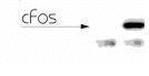
Western blot of cFos on NIH 3T3 cell lysate using Lane one shows non-immune serum and lane two shows immune serum.Application
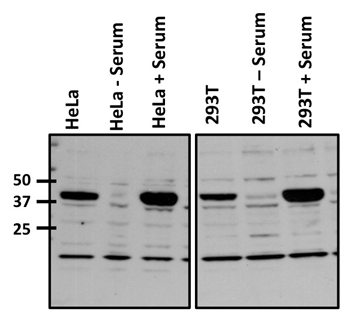
Western blot analysis of c-Fos was performed by loading 50ug of Serum (20% Serum, 2hours following 0.25% serum starvation, 36hours) treated HeLa and 293Twhole cell lysate, respectively, onto a 4-20% Tris-HCl polyacrylamide gel. Proteins were transferred to a PVDF membrane and blocked with 5% Milk/TBST for at least 1 hour. Membranes were probed with a rabbit polyclonal antibody recognizing c-Fos at a dilution of 1:500 overnight at 4°C on a rocking platform. Membranes were washed in TBS-0.1%Tween 20 and probed with a goat anti-rabbit-HRP secondary antibody at a dilution of 1:20,000 for at least one hour. Membranes were washed and chemiluminescent detection performed.Application
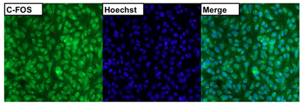
Immunofluorescent analysis of c-FOS (green) in untreated HeLa cells. Formalin fixed cells were permeabilized with 0.1% Triton X-100 in TBS for 15 minutes at room temperature. Cells were then blocked with 5% normal goat serum for 15 minutes at room temperature. Cells were probed with a rabbit polyclonal antibody recognizing c-FOS, at a dilution of 1:500 for at least 1 hour at room temperature. Cells were washed with PBS and incubated with DyLight 488 goat-anti-rabbit secondary antibody at a dilution of 1:400 for 30 minutes at room temperature. Nuclei were stained with Hoechst 33342 dye.| Product Name | c-Fos antibody |
|---|---|
| Antibody Type | Primary Antibodies |
| Product description | Rabbit Polyclonal antibody to c-FosCellular oncogenes, or proto-oncogenes, play pivotal roles in cellular communication pathways that regulate normal growth, development and differentiation. The cellular oncogene families fos and jun encode nuclear proteins that can function as transcription factors. The fos family of nuclear oncogenes encode cFos, FosB, (fos-related antigen) Fra1, and Fra2. Fos and Jun dimerize to form Activator Protein-1 (AP-1), a transcriptional factor that binds to the 12-O-tetradecanoylphorbol 13-acetate (TPA) response element (TRE) of several cellular and viral genes including human collagenase, metallothionein IIa, stromelysin, interleukin 2, SV40 and polyoma. Fos and Jun contain the ‘leucine-zipper’ motif that allows for dimerization and an adjacent basic domain required for biological activity. The functionally active form of Fos is in a heterodimer with a member of the Jun family. While Jun family members can form functional homodimers, studies indicate that Fos family members do not self-associate and therefore do not bind DNA on their own. The various dimers differ in their ability to transactivate AP-1 dependent genes. |
| Immunogen | Synthetic peptide corresponding to residues S(4) G F N A D Y E A S S S R C(17) of human cFos. |
| Clonality | Polyclonal |
|---|---|
| Host Species | Rabbit |
| Tested Applications | FACSICC/IFWB |
| ICC/IF: 1:500: |
|
| Species Reactivity | HumanMouse |
| Concentration | 1mg/ml (Please refer to the vial label for the specific concentration) |
| Purification | Affinity purified |
| Gene Synonyms | 2353 FOS 164810 c-fos cFos P01100 c fos |
|---|---|
| Target | c-Fos |
Application
Western blot of cFos on NIH 3T3 cell lysate using Lane one shows non-immune serum and lane two shows immune serum.
Application
Western blot analysis of c-Fos was performed by loading 50ug of Serum (20% Serum, 2hours following 0.25% serum starvation, 36hours) treated HeLa and 293Twhole cell lysate, respectively, onto a 4-20% Tris-HCl polyacrylamide gel. Proteins were transferred to a PVDF membrane and blocked with 5% Milk/TBST for at least 1 hour. Membranes were probed with a rabbit polyclonal antibody recognizing c-Fos at a dilution of 1:500 overnight at 4°C on a rocking platform. Membranes were washed in TBS-0.1%Tween 20 and probed with a goat anti-rabbit-HRP secondary antibody at a dilution of 1:20,000 for at least one hour. Membranes were washed and chemiluminescent detection performed.
Application
Immunofluorescent analysis of c-FOS (green) in untreated HeLa cells. Formalin fixed cells were permeabilized with 0.1% Triton X-100 in TBS for 15 minutes at room temperature. Cells were then blocked with 5% normal goat serum for 15 minutes at room temperature. Cells were probed with a rabbit polyclonal antibody recognizing c-FOS, at a dilution of 1:500 for at least 1 hour at room temperature. Cells were washed with PBS and incubated with DyLight 488 goat-anti-rabbit secondary antibody at a dilution of 1:400 for 30 minutes at room temperature. Nuclei were stained with Hoechst 33342 dye.| Application Notes | ICC/IF: 1:500: |
|---|
| Form | Liquid |
|---|---|
| Storage Instructions | After reconstitution, keep as concentrated solution. Aliquot and store at -20ºC or below. Avoid multiple freeze-thaw cycles. |
| Storage Buffer | Lyophilized, purified rabbit IgG containing 0.05% sodium azide. Reconstitute with 200 ul PBS. |


