Application
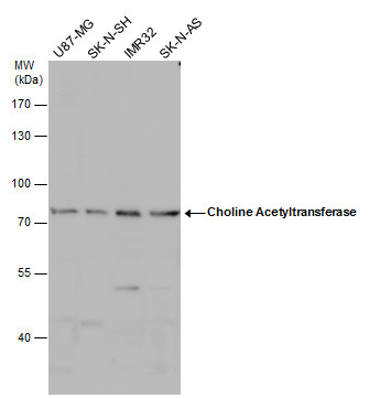
Choline Acetyltransferase antibody [N1N3] detects Choline Acetyltransferase protein by western blot analysis. Various whole cell extracts (30 µg) were separated by 7.5% SDS-PAGE, and the membrane was blotted with Choline Acetyltransferase antibody [N1N3] diluted at 1:500. The HRP-conjugated anti-rabbit IgG antibody was used to detect the primary antibody.Application
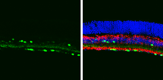
Choline Acetyltransferase antibody [N1N3] detects Choline Acetyltransferase protein in the amacrine cells by immunohistochemical analysis. Sample: Frozen sectioned adult mouse retina. Green: Choline Acetyltransferase protein stained by Choline Acetyltransferase antibody [N1N3] diluted at 1:250. Red: Protein kinase C alpha staining. Blue: Fluoroshield with DAPI .Application
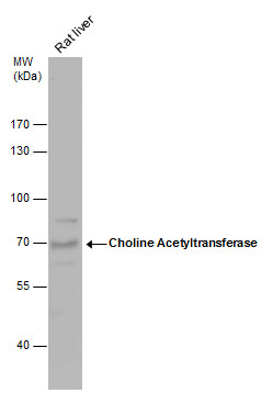
Choline Acetyltransferase antibody [N1N3] detects Choline Acetyltransferase protein by western blot analysis. Rat tissue extracts (50 µg) was separated by 7.5% SDS-PAGE, and the membrane was blotted with Choline Acetyltransferase antibody [N1N3] diluted at 1:500. The HRP-conjugated anti-rabbit IgG antibody was used to detect the primary antibody.Application
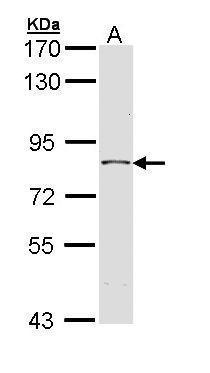
Sample (30 µg of whole cell lysate) A: H1299 7.5% SDS PAGE diluted at 1:1000 The HRP-conjugated anti-rabbit IgG antibody was used to detect the primary antibody.Application
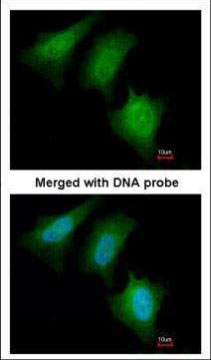
Immunofluorescence analysis of paraformaldehyde-fixed HeLa, using CHAT antibodyApplication
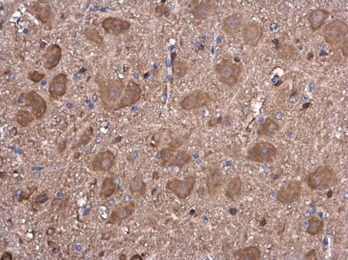
Choline Acetyltransferase antibody [N1N3] detects Choline Acetyltransferase protein at cytoplasm in mouse brain by immunohistochemical analysis. Sample: Paraffin-embedded mouse brain. Choline Acetyltransferase antibody [N1N3] diluted at 1:100.IHC
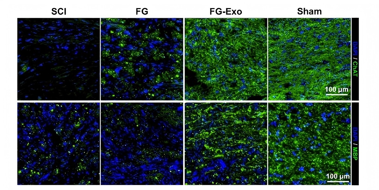
Immunostaining of the injured spinal cord tissues for choline acetyltransferase (ChAT, green) and myelin basic protein (MBP, green) in SCI, FG, FG-Exo, and Sham groups. Nuclei were stained with DAPI (blue).| Product Name | Choline Acetyltransferase antibody |
|---|---|
| Antibody Type | Primary Antibodies |
| Product description | Rabbit polyclonal antibody to CHAT (choline acetyltransferase)This gene encodes an enzyme which catalyzes the biosynthesis of the neurotransmitter acetylcholine. This gene product is a characteristic feature of cholinergic neurons, and changes in these neurons may explain some of the symptoms of Alzheimer disease. Mutations in this gene are associated with congenital myasthenic syndrome associated with episodic apnea. Multiple transcript variants encoding different isoforms have been found for this gene, and some of these variants have been shown to encode more than one isoform. [provided by RefSeq] |
| Immunogen | Recombinant protein encompassing a sequence within the N-terminus region of human Choline Acetyltransferase. The exact sequence is proprietary. |
| Isotype | IgG |
|---|---|
| Host Species | Rabbit |
| Tested Applications | ICC/IFIHCIHC-FIHC-PWB |
| WB:1:500~1:1000 ICC/IF:1:100~1:500 IHC:1:50~1:100 *Optimal dilutions/concentrations should be determined by the researcher. |
|
| Species Reactivity | HumanMouseRat |
| Predicted Reactivity | PigPig (94%) |
| Concentration | 1mg/ |
| Purification | Affinity purified |
| Gene Synonyms | CHOACTASE antibody, CMS1A antibody, CMS1A2 antibody, CHAT antibody, choline O-acetyltransferase antibody, acetyl CoA:choline O-acetyltransferase antibody, choline acetylase antibody |
|---|---|
| Target | CHAT |
| Molecular Weight(MW) | 83 kDa (note) (8) |
Application
Choline Acetyltransferase antibody [N1N3] detects Choline Acetyltransferase protein by western blot analysis. Various whole cell extracts (30 µg) were separated by 7.5% SDS-PAGE, and the membrane was blotted with Choline Acetyltransferase antibody [N1N3] diluted at 1:500. The HRP-conjugated anti-rabbit IgG antibody was used to detect the primary antibody.
Application
Choline Acetyltransferase antibody [N1N3] detects Choline Acetyltransferase protein in the amacrine cells by immunohistochemical analysis. Sample: Frozen sectioned adult mouse retina. Green: Choline Acetyltransferase protein stained by Choline Acetyltransferase antibody [N1N3] diluted at 1:250. Red: Protein kinase C alpha staining. Blue: Fluoroshield with DAPI .
Application
Choline Acetyltransferase antibody [N1N3] detects Choline Acetyltransferase protein by western blot analysis. Rat tissue extracts (50 µg) was separated by 7.5% SDS-PAGE, and the membrane was blotted with Choline Acetyltransferase antibody [N1N3] diluted at 1:500. The HRP-conjugated anti-rabbit IgG antibody was used to detect the primary antibody.
Application
Sample (30 µg of whole cell lysate) A: H1299 7.5% SDS PAGE diluted at 1:1000 The HRP-conjugated anti-rabbit IgG antibody was used to detect the primary antibody.
Application
Immunofluorescence analysis of paraformaldehyde-fixed HeLa, using CHAT antibody
Application
Choline Acetyltransferase antibody [N1N3] detects Choline Acetyltransferase protein at cytoplasm in mouse brain by immunohistochemical analysis. Sample: Paraffin-embedded mouse brain. Choline Acetyltransferase antibody [N1N3] diluted at 1:100.
IHC
Immunostaining of the injured spinal cord tissues for choline acetyltransferase (ChAT, green) and myelin basic protein (MBP, green) in SCI, FG, FG-Exo, and Sham groups. Nuclei were stained with DAPI (blue).| Positive Control | H1299 , U87-MG , SK-N-SH , IMR32 , SK-N-AS , Mouse brain , Rat liver |
|---|---|
| Application Notes | WB:1:500~1:1000 ICC/IF:1:100~1:500 IHC:1:50~1:100 *Optimal dilutions/concentrations should be determined by the researcher. |
| Form | Liquid |
|---|---|
| Storage Instructions | Keep as concentrated solution. Aliquot and store at -20ºC or below. Avoid multiple freeze-thaw cycles. |
| Storage Buffer | 0.1M Tris, 0.1M Glycine, 10% Glycerol (pH7). 0.01% Thimerosal was added as a preservative. |


