Application
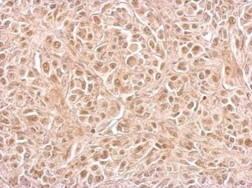
CIAPIN1 antibody detects CIAPIN1 protein at cytosol and nucleus on SkHep1 xenograft by immunohistochemical analysis. Sample: Paraffin-embedded SkHep1 xenograft. CIAPIN1 antibody dilution: 1:500.Application
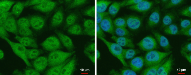
CIAPIN1 antibody detects CIAPIN1 protein at nucleus and cytoplasm by immunofluorescent analysis. Sample: A375 cells were fixed in 4% paraformaldehyde at RT for 15 min. Green: CIAPIN1 protein stained by CIAPIN1 antibody diluted at 1:500. Blue: Hoechst 33342 staining. Scale bar = 10 μm.Application
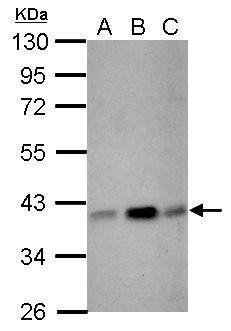
Sample (30 ug of whole cell lysate) A: NIH-3T3 B: JC C: BCL-1 10% SDS PAGE diluted at 1:3000Application
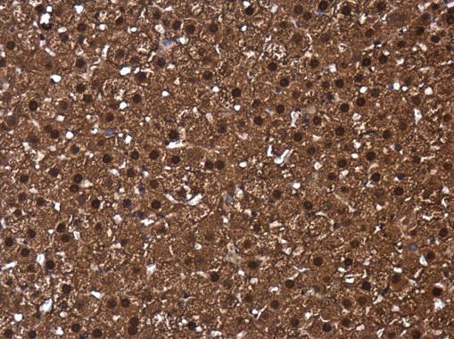
CIAPIN1 antibody detects CIAPIN1 protein at cytoplasm and nucleus in rat adrenal gland by immunohistochemical analysis. Sample: Paraffin-embedded rat adrenal gland. CIAPIN1 antibody diluted at 1:500.Application
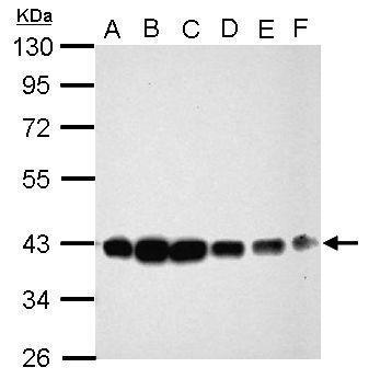
Sample (30 ug of whole cell lysate) A: Jurkat B: Raji C: K562 D: THP-1 E: HL-60 F: NCI-H929 10% SDS PAGE diluted at 1:1000| Product Name | CIAPIN1 antibody |
|---|---|
| Antibody Type | Primary Antibodies |
| Product description | Rabbit Polyclonal antibody to CIAPIN1 (cytokine induced apoptosis inhibitor 1)CIAPIN1 is a cytokine-induced inhibitor of apoptosis with no relation to apoptosis regulatory molecules of the BCL2 (MIM 151430) or CASP (see MIM 147678) families. Expression of CIAPIN1 is dependent on growth factor stimulation (Shibayama et al., 2004 [PubMed 14970183]).[supplied by OMIM] |
| Immunogen | Full length human CIAPIN1 Recombinant protein. |
| Clonality | Polyclonal |
|---|---|
| Isotype | IgG |
| Host Species | Rabbit |
| Tested Applications | ICC/IFIHC-PWB |
| *Optimal dilutions/concentrations should be determined by the researcher. | |
| Species Reactivity | HumanMouseRat |
| Predicted Reactivity | Chimpanzee, BovineChimpanzee (99%), Bovine (90%) |
| Concentration | 1mg/ml (Please refer to the vial label for the specific concentration) |
| Purification | Affinity purified |
| Gene Synonyms | 2810413N20Rik antibody, Anamorsin antibody, DRE2 antibody, PRO0915 antibody, CIAPIN1 antibody, predicted protein of HQ0915 antibody, fe-S cluster assembly protein DRE2 homolog antibody, cytokine-induced apoptosis inhibitor 1 antibody, cytokine induced apop |
|---|---|
| Molecular Weight(MW) | 34 kDa (note) (2) |
| Cellular Localization | Cytoplasm |
Application
CIAPIN1 antibody detects CIAPIN1 protein at cytosol and nucleus on SkHep1 xenograft by immunohistochemical analysis. Sample: Paraffin-embedded SkHep1 xenograft. CIAPIN1 antibody dilution: 1:500.
Application
CIAPIN1 antibody detects CIAPIN1 protein at nucleus and cytoplasm by immunofluorescent analysis. Sample: A375 cells were fixed in 4% paraformaldehyde at RT for 15 min. Green: CIAPIN1 protein stained by CIAPIN1 antibody diluted at 1:500. Blue: Hoechst 33342 staining. Scale bar = 10 μm.
Application
Sample (30 ug of whole cell lysate) A: NIH-3T3 B: JC C: BCL-1 10% SDS PAGE diluted at 1:3000
Application
CIAPIN1 antibody detects CIAPIN1 protein at cytoplasm and nucleus in rat adrenal gland by immunohistochemical analysis. Sample: Paraffin-embedded rat adrenal gland. CIAPIN1 antibody diluted at 1:500.
Application
Sample (30 ug of whole cell lysate) A: Jurkat B: Raji C: K562 D: THP-1 E: HL-60 F: NCI-H929 10% SDS PAGE diluted at 1:1000| Positive Control | Jurkat , Raji , K562 , THP-1 , HL-60 , NCI-H929 , NIH-3T3 , JC , BCL-1 |
|---|---|
| Application Notes | *Optimal dilutions/concentrations should be determined by the researcher. |
| Form | Liquid |
|---|---|
| Storage Instructions | Keep as concentrated solution. Aliquot and store at -20ºC or below. Avoid multiple freeze-thaw cycles. |
| Storage Buffer | 1XPBS, 20% Glycerol (pH7). 0.01% Thimerosal was added as a preservative. |


