Application
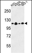
Western blot analysis of COL17A1 Antibody (Center) in A375, A2058, HL-60, MCF-7 cell line lysates (35ug/lane). COL17A1 (arrow) was detected using the purified Pab.Application
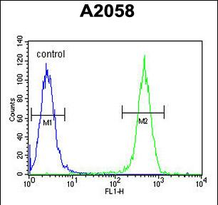
COL17A1 Antibody (Center) flow cytometric analysis of A2058 cells (right histogram) compared to a negative control cell (left histogram). FITC-conjugated goat-anti-rabbit secondary antibodies were used for the analysis.Application
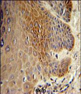
COL17A1 Antibody (Center) IHC analysis in formalin fixed and paraffin embedded skin tissue followed by peroxidase conjugation of the secondary antibody and DAB staining. This data demonstrates the use of the COL17A1 Antibody (Center) for immunohistochemistry.| Product Name | COL17A1 antibody, Internal |
|---|---|
| Antibody Type | Primary Antibodies |
| Product description | Rabbit polyclonal to COL17A1 (Internal)This gene encodes the alpha chain of type XVII collagen. Unlike most collagens, collagen XVII is a transmembrane protein. Collagen XVII is a structural component of hemidesmosomes, multiprotein complexes at the dermal-epidermal basement membrane zone that mediate adhesion of keratinocytes to the underlying membrane. Mutations in this gene are associated with both generalized atrophic benign and junctional epidermolysis bullosa. Two homotrimeric forms of type XVII collagen exist. The full length form is the transmembrane protein. A soluble form, referred to as either ectodomain or LAD-1, is generated by proteolytic processing of the full length form. [provided by RefSeq] |
| Immunogen | The immunogenic sequence of GTX80563 is between amino acids 490-520 of human COL17A1 |
| Clonality | Polyclonal |
|---|---|
| Isotype | IgG |
| Host Species | Rabbit |
| Tested Applications | FACSIHC-PWB |
| ELISA: Use at 1:1,000: |
|
| Species Reactivity | Human |
| Concentration | 0.2mg/ml |
| Purification | Affinity purified |
| Gene Synonyms | 97 kDa linear IgA disease antigen Linear IgA disease antigen 1 LAD-1 120 kDa linear IgA disease antigen Linear IgA disease antigen1 Linear IgA disease antigen 1 120 kDa linear IgA dermatosis antigen 97 kDa linear IgA bullous dermatosis antigen 97 |
|---|---|
| Target | COL17A1 |
| Molecular Weight(MW) | 150.419 kDa (note) |
| Cellular Localization | Localized along the plasma membrane of the hemidesmosome. Exclusively localized to anchoring filaments. Localized to the epidermal side of split skin. Localized in the lamina lucida beneath the hemidesmosomes. |
Application
Western blot analysis of COL17A1 Antibody (Center) in A375, A2058, HL-60, MCF-7 cell line lysates (35ug/lane). COL17A1 (arrow) was detected using the purified Pab.
Application
COL17A1 Antibody (Center) flow cytometric analysis of A2058 cells (right histogram) compared to a negative control cell (left histogram). FITC-conjugated goat-anti-rabbit secondary antibodies were used for the analysis.
Application
COL17A1 Antibody (Center) IHC analysis in formalin fixed and paraffin embedded skin tissue followed by peroxidase conjugation of the secondary antibody and DAB staining. This data demonstrates the use of the COL17A1 Antibody (Center) for immunohistochemistry.| Application Notes | ELISA: Use at 1:1,000: |
|---|
| Form | Liquid |
|---|---|
| Storage Instructions | Store at at 2-8°C for up to 6 months. For long term storage, aliquot and store at -20 °C. Avoid repeated freeze/thaw cycles. |
| Storage Buffer | PBS with 0.09% (W/V) sodium azide |


