Application
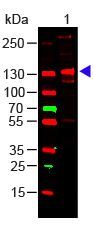
Western Blot of Rabbit Anti-Collagen III Antibody Load: 100 ng Human Collagen III. Primary antibody: Collagen III Antibody at 1:1000 o/n at 4°C. Secondary antibody: DyLight™ 649 Goat anti-rabbit at 1:20,000 for 30 min at RT Block for 30 min at RT. Predicted/Observed size: 138 kDa, 138 kDa.Application
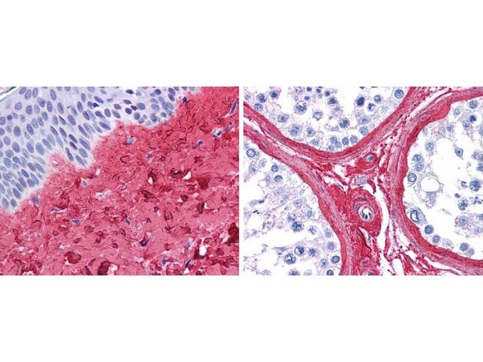
GeneTex anti collagen III antibody ( 45 min RT) showed strong staining in FFPE sections of human skin(left, dermis) with moderate to strong red staining and testis (right) where strong staining was observed within connective tissue between seminiferous tubules. The antibody showed strong extracellular staining within connective tissues across many organs with minimal background staining. Slides were steamed in 0.01 M sodium citrate buffer, pH 6.0 at 99-100°C - 20 minutes for antigen retrieval.| Product Name | Collagen III antibody |
|---|---|
| Antibody Type | Primary Antibodies |
| Product description | Rabbit Polyclonal antibody to Collagen IIIThese antibodies are well suited to detect extracellular matrix proteins in normal as well as disease state tissues. Disruption of tissue organization is the hallmark of neoplasia. Malignant lesions can be distinguished from benign by examining the breakdown of basement membranes and loss of 3-dimensional architecture. Malignant cells are presumed to use matrix metalloproteases to degrade barriers created by the extracellular matrix, which then allows metastasis to occur. Collagenases, stomelysins and gelatinases can collectively degrade all of the various components of the extracellular matrix, including fibrillar and non-fibrillar collagens and basement membrane glycoproteins. |
| Immunogen | Collagen Type III from human and bovine placenta |
| Clonality | Polyclonal |
|---|---|
| Isotype | IgG |
| Host Species | Rabbit |
| Tested Applications | ELISAIHC-FIHC-PIPWB |
| *Optimal dilutions/concentrations should be determined by the researcher.
: |
|
| Species Reactivity | HumanBovine |
| Concentration | 1mg/ml |
| Purification | Affinity purified |
| Gene Synonyms | COL3A1 EDS4A |
|---|---|
| Target | Collagen III |
| Cellular Localization | Extracellular matrix |
Application
Western Blot of Rabbit Anti-Collagen III Antibody Load: 100 ng Human Collagen III. Primary antibody: Collagen III Antibody at 1:1000 o/n at 4°C. Secondary antibody: DyLight™ 649 Goat anti-rabbit at 1:20,000 for 30 min at RT Block for 30 min at RT. Predicted/Observed size: 138 kDa, 138 kDa.
Application
GeneTex anti collagen III antibody ( 45 min RT) showed strong staining in FFPE sections of human skin(left, dermis) with moderate to strong red staining and testis (right) where strong staining was observed within connective tissue between seminiferous tubules. The antibody showed strong extracellular staining within connective tissues across many organs with minimal background staining. Slides were steamed in 0.01 M sodium citrate buffer, pH 6.0 at 99-100°C - 20 minutes for antigen retrieval.| Application Notes | *Optimal dilutions/concentrations should be determined by the researcher.
: |
|---|
| Form | Liquid |
|---|---|
| Storage Instructions | For extended storage, mix with an equal volume of glycerol, aliquot contents and freeze at -20° C or below. Avoid cycles of freezing and thawing. |
| Storage Buffer | 0.125 M Sodium Borate, 0.075 M Sodium Chloride, 0.005 M EDTA, pH 8.0 and 0.01% (w/v) Sodium Azide |


