Application
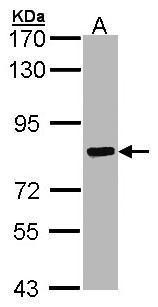
Sample (30 ug of whole cell lysate) A: Hep G2 7.5% SDS PAGE diluted at 1:1000Application
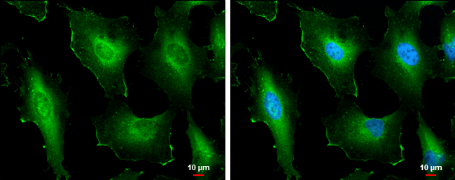
Cortactin antibody [N1], N-term detects Cortactin protein at cell membrane and cytoplasm by immunofluorescent analysis. Sample: HeLa cells were fixed in 4% paraformaldehyde at RT for 15 min. Green: Cortactin protein stained by Cortactin antibody [N1], N-term diluted at 1:500. Blue: Hoechst 33342 staining. Scale bar = 10 μm.Application
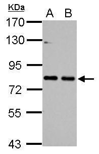
Sample (30 ug of whole cell lysate) A: NIH-3T3 B: JC 7.5% SDS PAGE diluted at 1:1000Application
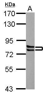
Sample (30 ug of whole cell lysate) A: PC-12 7.5% SDS PAGE diluted at 1:3000Application
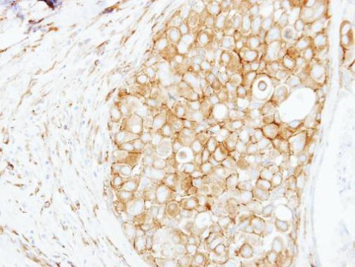
Immunohistochemical analysis of paraffin-embedded human breast cancer, using Cortactin antibody at 1:250 dilution.Application
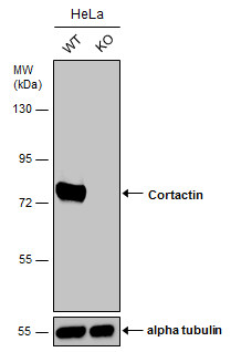
Wild-type (WT) and Cortactin knockout (KO) HeLa cell extracts (30 µg) were separated by 7.5% SDS-PAGE, and the membrane was blotted with Cortactin antibody [N1], N-term diluted at 1:1000. The HRP-conjugated anti-rabbit IgG antibody was used to detect the primary antibody.| Product Name | Cortactin antibody , N-term |
|---|---|
| Antibody Type | Primary Antibodies |
| Product description | Rabbit Polyclonal antibody to Cortactin (src substrate cortactin)This gene is overexpressed in breast cancer and squamous cell carcinomas of the head and neck. The encoded protein is localized in the cytoplasm and in areas of the cell-substratum contacts. This gene has two roles: (1) regulating the interactions between components of adherens-type junctions and (2) organizing the cytoskeleton and cell adhesion structures of epithelia and carcinoma cells. During apoptosis, the encoded protein is degraded in a caspase-dependent manner. The aberrant regulation of this gene contributes to tumor cell invasion and metastasis. Two splice variants that encode different isoforms have been identified for this gene. [provided by RefSeq] |
| Immunogen | Carrier-protein conjugated synthetic peptide encompassing a sequence within the N-terminus region of human Cortactin. The exact sequence is proprietary. |
| Clonality | Polyclonal |
|---|---|
| Isotype | IgG |
| Host Species | Rabbit |
| Tested Applications | ICC/IFIHC-PWB |
| *Optimal dilutions/concentrations should be determined by the researcher.
: |
|
| Species Reactivity | HumanMouseRat |
| Predicted Reactivity | Rhesus MonkeyRhesus Monkey (100%) |
| Concentration | 1mg/ml (Please refer to the vial label for the specific concentration) |
| Purification | Affinity purified |
| Gene Synonyms | EMS1 antibody FLJ34459 antibody CTTN antibody oncogene EMS1 antibody src substrate cortactin antibody amplaxin antibody ems1 sequence (mammary tumor and squamous cell carcinoma-associated (p80/85 src substrate) antibody 1110020L01Rik antibody corta |
|---|---|
| Molecular Weight(MW) | 62 kDa (note) (3) |
| Cellular Localization | Cytoplasm , cytoskeleton (By similarity) , Cell projection , lamellipodium (By similarity) , ruffle (By similarity) |
Application
Sample (30 ug of whole cell lysate) A: Hep G2 7.5% SDS PAGE diluted at 1:1000
Application
Cortactin antibody [N1], N-term detects Cortactin protein at cell membrane and cytoplasm by immunofluorescent analysis. Sample: HeLa cells were fixed in 4% paraformaldehyde at RT for 15 min. Green: Cortactin protein stained by Cortactin antibody [N1], N-term diluted at 1:500. Blue: Hoechst 33342 staining. Scale bar = 10 μm.
Application
Sample (30 ug of whole cell lysate) A: NIH-3T3 B: JC 7.5% SDS PAGE diluted at 1:1000
Application
Sample (30 ug of whole cell lysate) A: PC-12 7.5% SDS PAGE diluted at 1:3000
Application
Immunohistochemical analysis of paraffin-embedded human breast cancer, using Cortactin antibody at 1:250 dilution.
Application
Wild-type (WT) and Cortactin knockout (KO) HeLa cell extracts (30 µg) were separated by 7.5% SDS-PAGE, and the membrane was blotted with Cortactin antibody [N1], N-term diluted at 1:1000. The HRP-conjugated anti-rabbit IgG antibody was used to detect the primary antibody.| Positive Control | HepG2 , NIH-3T3 , JC , PC-12 |
|---|---|
| Application Notes | *Optimal dilutions/concentrations should be determined by the researcher.
: |
| Form | Liquid |
|---|---|
| Storage Instructions | Keep as concentrated solution. Aliquot and store at -20ºC or below. Avoid multiple freeze-thaw cycles. |
| Storage Buffer | 1XPBS, 1% BSA, 20% Glycerol (pH7). 0.01% Thimerosal was added as a preservative. |


