Application
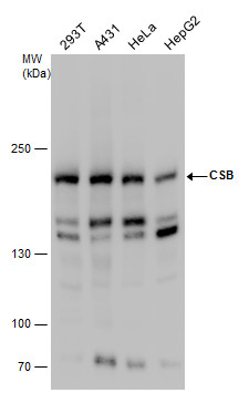
CSB antibody detects CSB protein by western blot analysis. Various whole cell extracts (30 µg) were separated by 5% SDS-PAGE, and the membrane was blotted with CSB antibody diluted at a dilution of 1:500. The HRP-conjugated anti-rabbit IgG antibody was used to detect the primary antibody.Application
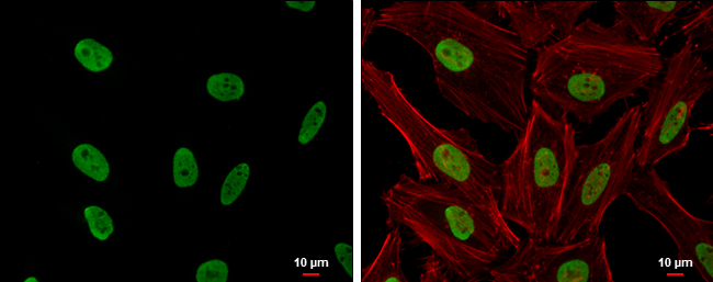
CSB antibody [N2C1], Internal detects CSB protein at nucleus by immunofluorescent analysis. Sample: HeLa cells were fixed in 4% paraformaldehyde at RT for 15 min. Green: CSB protein stained by CSB antibody [N2C1], Internal diluted at 1:500. Red: phalloidin, a cytoskeleton marker, diluted at 1:200. Blue: Hoechst 33342 staining. Scale bar = 10 μm.Application
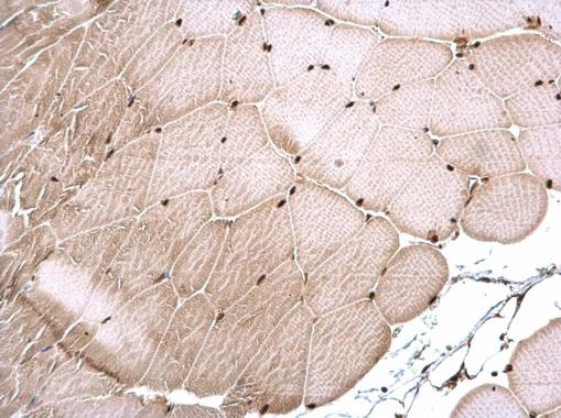
CSB antibody [N2C1], Internal detects CSB protein at nucleus on mouse muscle by immunohistochemical analysis. Sample: Paraffin-embedded mouse muscle. CSB antibody [N2C1], Internal dilution: 1:500.Application
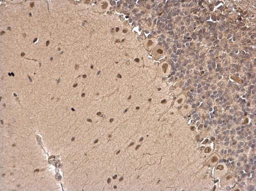
CSB antibody [N2C1], Internal detects CSB protein at nucleus on rat hind brain by immunohistochemical analysis. Sample: Paraffin-embedded rat hind brain. CSB antibody [N2C1], Internal dilution: 1:500.Application
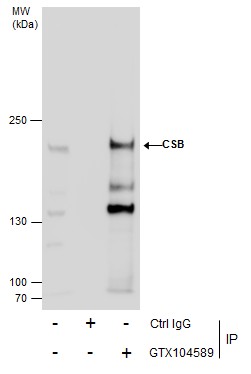
Immunoprecipitation of CSB protein from 293T whole cell extracts using 5 μg of CSB antibody [N2C1], Internal . Western blot analysis was performed using CSB antibody [N2C1], Internal . EasyBlot anti-Rabbit IgG was used as a secondary reagent.| Product Name | CSB antibody , Internal |
|---|---|
| Antibody Type | Primary Antibodies |
| Product description | Rabbit Polyclonal antibody to CSB (excision repair cross-complementing rodent repair deficiency, complementation group 6)This gene encodes a DNA-binding protein that is important in transcription-coupled excision repair. The protein has ATP-stimulated ATPase activity; there are contradictory publications reporting presence or absence of helicase activity. The protein appears to interact with several transcription and excision repair proteins, and may promote complex formation at repair sites. Mutations in this gene result in Cockayne syndrome type B. [provided by RefSeq] |
| Immunogen | Recombinant protein encompassing a sequence within the center region of human CSB. The exact sequence is proprietary. |
| Clonality | Polyclonal |
|---|---|
| Isotype | IgG |
| Host Species | Rabbit |
| Tested Applications | ICC/IFIHC-PIPWB |
| *Optimal dilutions/concentrations should be determined by the researcher.
: |
|
| Species Reactivity | HumanMouseRat |
| Predicted Reactivity | BovineBovine (90%) |
| Concentration | 1mg/ml |
| Purification | Affinity purified |
| Gene Synonyms | ARMD5 antibody CKN2 antibody COFS antibody COFS1 antibody CSB antibody RAD26 antibody ERCC6 antibody Cockayne syndrome B protein antibody ATP-dependent helicase ERCC6 antibody cockayne syndrome protein CSB antibody Rad26 homolog antibody Cockayn |
|---|---|
| Molecular Weight(MW) | 168 kDa (note) |
| Cellular Localization | Nucleus |
Application
CSB antibody detects CSB protein by western blot analysis. Various whole cell extracts (30 µg) were separated by 5% SDS-PAGE, and the membrane was blotted with CSB antibody diluted at a dilution of 1:500. The HRP-conjugated anti-rabbit IgG antibody was used to detect the primary antibody.
Application
CSB antibody [N2C1], Internal detects CSB protein at nucleus by immunofluorescent analysis. Sample: HeLa cells were fixed in 4% paraformaldehyde at RT for 15 min. Green: CSB protein stained by CSB antibody [N2C1], Internal diluted at 1:500. Red: phalloidin, a cytoskeleton marker, diluted at 1:200. Blue: Hoechst 33342 staining. Scale bar = 10 μm.
Application
CSB antibody [N2C1], Internal detects CSB protein at nucleus on mouse muscle by immunohistochemical analysis. Sample: Paraffin-embedded mouse muscle. CSB antibody [N2C1], Internal dilution: 1:500.
Application
CSB antibody [N2C1], Internal detects CSB protein at nucleus on rat hind brain by immunohistochemical analysis. Sample: Paraffin-embedded rat hind brain. CSB antibody [N2C1], Internal dilution: 1:500.
Application
Immunoprecipitation of CSB protein from 293T whole cell extracts using 5 μg of CSB antibody [N2C1], Internal . Western blot analysis was performed using CSB antibody [N2C1], Internal . EasyBlot anti-Rabbit IgG was used as a secondary reagent.| Positive Control | 293T , A431 , HeLa , HepG2 |
|---|---|
| Application Notes | *Optimal dilutions/concentrations should be determined by the researcher.
: |
| Form | Liquid |
|---|---|
| Storage Instructions | Keep as concentrated solution. Aliquot and store at -20ºC or below. Avoid multiple freeze-thaw cycles. |
| Storage Buffer | 1XPBS, 0.2M Tris, 0.2M Glycine, 1mM EGTA, 50% Glycerol (pH7). 0.025% ProClin 300 was added as a preservative. |


