Application
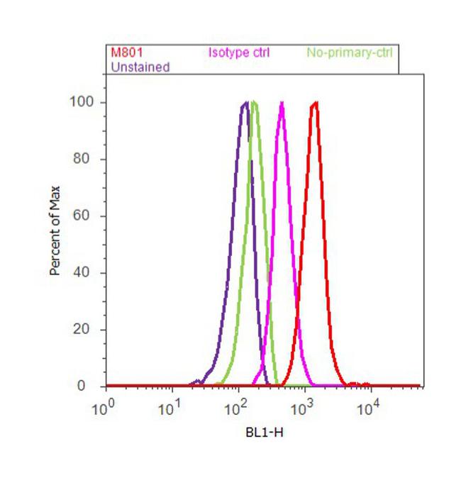
Flow cytometry analysis of IL-8 was done on U-87 MG cells. Cells were fixed with 70% ethanol for 10 minutes, permeabilized with 0.25% Triton™ X-100 for 20 minutes, and blocked with 5% BSA for 30 minutes at room temperature. Cells were labeled with IL-8 Mouse Monoclonal Antibody (M801, red histogram) or with mouse isotype control (pink histogram) at 3-5 µg/million cells in 2.5% BSA. After incubation at room temperature for 2 hours, the cells were labeled with Alexa Fluor® 488 Rabbit Anti-Mouse Secondary Antibody at a dilution of 1:400 for 30 minutes at room temperature. The representative 10,000 cells were acquired and analyzed for each sample using an Attune® Acoustic Focusing Cytometer. The purple histogram represents unstained control cells and the green histogram represents no-primary-antibody control.Application
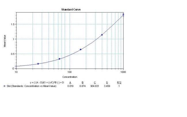
Sandwich ELISA analysis of human IL-8 was performed using a Human IL-8 Colorimetric ELISA kit by loading 50 ul per well of Human IL-8 Recombinant Protein in dodecuplicate at 1000, 400, 160, 64, 25.6, and 0 pg/ml across a 3 ug/ml mouse anti-Human IL-8 (M801) pre-coated plate and incubating for 1 hour at room temperature. The plate was washed, and then incubated with 50 ul per well of a biotinylated mouse anti-human IL-8 monoclonal antibody in duplicate at 0.25 ug/ml for 1 hour at room temperature. The plate was washed and incubated with 100 ul per well of Streptavidin-HRP in all test wells at a 1:4,000 dilution for 30 minutes at room temperature. Detection was performed using 1-Step Ultra TMB substrate for 30 minutes at room temperature in the dark. The plate was then stopped with 0.16M sulfuric acid. Absorbances were read on a spectrophotometer at 450-550 nm.Application
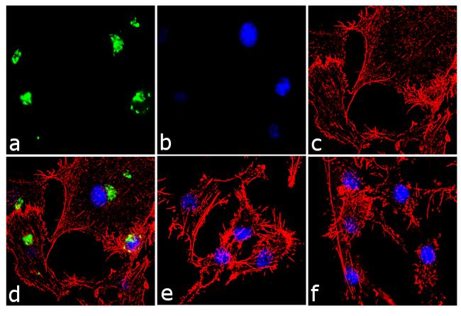
Immunofluorescence analysis of IL-8 was performed using 70% confluent log phase U-87 MG cells treated 1 uM thapsigargin for 24 hours. The cells were fixed with 4% paraformaldehyde for 10 minutes, permeabilized with 0.1% Triton™ X-100 for 10 minutes, and blocked with 1% BSA for 1 hour at room temperature. The cells were labeled with IL-8 Mouse Monoclonal Antibody (M801) at 2µg/ml in 0.1% BSA and incubated for 3 hours at room temperature and then labeled with Goat anti-Mouse IgG (H+L) Superclonal™ Secondary Antibody, Alexa Fluor® 488 conjµgate at a dilution of 1:2000 for 45 minutes at room temperature (Panel a: green). Nuclei (Panel b: blue) were stained with SlowFade® Gold Antifade Mountant with DAPI. F-actin (Panel c: red) was stained with Alexa Fluor® 555 Rhodamine Phalloidin. Panel d represents the merged image showing cytoplasmic localization. Panel e is untreated cell with no signal. Panel f represents control cells with no primary antibody to assess background. The images were captured at 60X magnification..| Product Name | CXCL8 / IL8 antibody |
|---|---|
| Antibody Type | Primary Antibodies |
| Product description | Mouse monoclonal antibody [3IL8-H10] to CXCL8The protein encoded by this gene is a member of the CXC chemokine family. This chemokine is one of the major mediators of the inflammatory response. This chemokine is secreted by several cell types. It functions as a chemoattractant, and is also a potent angiogenic factor. This gene is believed to play a role in the pathogenesis of bronchiolitis, a common respiratory tract disease caused by viral infection. This gene and other ten members of the CXC chemokine gene family form a chemokine gene cluster in a region mapped to chromosome 4q. [provided by RefSeq, Jul 2008] |
| Immunogen | Recombinant human IL-8 |
| Clonality | Monoclonal |
|---|---|
| Isotype | IgG |
| Clone Number | 3IL8-H10 |
| Host Species | Mouse |
| Tested Applications | ELISAIHCWB |
| IHC:1:50~1:200 WB:1:500~1:2000 Notes:Optimal dilutions/concentrations should be determined by the researcher. |
|
| Species Reactivity | HumanMouse |
| Concentration | 1mg/ml |
| Purification | Protein A |
| Gene Synonyms | GCP-1 MDNCF NAP-1 LYNAP NAP1 MONAP GCP1 interleukin 8 CXCL8 LUCT LECT IL8 NAF |
|---|---|
| Target | IL-8 |
Application
Flow cytometry analysis of IL-8 was done on U-87 MG cells. Cells were fixed with 70% ethanol for 10 minutes, permeabilized with 0.25% Triton™ X-100 for 20 minutes, and blocked with 5% BSA for 30 minutes at room temperature. Cells were labeled with IL-8 Mouse Monoclonal Antibody (M801, red histogram) or with mouse isotype control (pink histogram) at 3-5 µg/million cells in 2.5% BSA. After incubation at room temperature for 2 hours, the cells were labeled with Alexa Fluor® 488 Rabbit Anti-Mouse Secondary Antibody at a dilution of 1:400 for 30 minutes at room temperature. The representative 10,000 cells were acquired and analyzed for each sample using an Attune® Acoustic Focusing Cytometer. The purple histogram represents unstained control cells and the green histogram represents no-primary-antibody control.
Application
Sandwich ELISA analysis of human IL-8 was performed using a Human IL-8 Colorimetric ELISA kit by loading 50 ul per well of Human IL-8 Recombinant Protein in dodecuplicate at 1000, 400, 160, 64, 25.6, and 0 pg/ml across a 3 ug/ml mouse anti-Human IL-8 (M801) pre-coated plate and incubating for 1 hour at room temperature. The plate was washed, and then incubated with 50 ul per well of a biotinylated mouse anti-human IL-8 monoclonal antibody in duplicate at 0.25 ug/ml for 1 hour at room temperature. The plate was washed and incubated with 100 ul per well of Streptavidin-HRP in all test wells at a 1:4,000 dilution for 30 minutes at room temperature. Detection was performed using 1-Step Ultra TMB substrate for 30 minutes at room temperature in the dark. The plate was then stopped with 0.16M sulfuric acid. Absorbances were read on a spectrophotometer at 450-550 nm.
Application
Immunofluorescence analysis of IL-8 was performed using 70% confluent log phase U-87 MG cells treated 1 uM thapsigargin for 24 hours. The cells were fixed with 4% paraformaldehyde for 10 minutes, permeabilized with 0.1% Triton™ X-100 for 10 minutes, and blocked with 1% BSA for 1 hour at room temperature. The cells were labeled with IL-8 Mouse Monoclonal Antibody (M801) at 2µg/ml in 0.1% BSA and incubated for 3 hours at room temperature and then labeled with Goat anti-Mouse IgG (H+L) Superclonal™ Secondary Antibody, Alexa Fluor® 488 conjµgate at a dilution of 1:2000 for 45 minutes at room temperature (Panel a: green). Nuclei (Panel b: blue) were stained with SlowFade® Gold Antifade Mountant with DAPI. F-actin (Panel c: red) was stained with Alexa Fluor® 555 Rhodamine Phalloidin. Panel d represents the merged image showing cytoplasmic localization. Panel e is untreated cell with no signal. Panel f represents control cells with no primary antibody to assess background. The images were captured at 60X magnification..| Application Notes | IHC:1:50~1:200 WB:1:500~1:2000 Notes:Optimal dilutions/concentrations should be determined by the researcher. |
|---|
| Form | Liquid |
|---|---|
| Storage Instructions | -20°C |
| Storage Buffer | PBS |


