Application
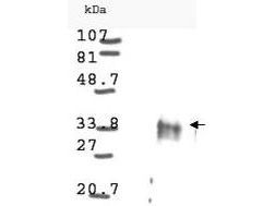
Western blot analysis is shown using GeneTex Anti-Cyclin D1 antibody to detect Human Cyclin D1 present in asynchronous HN30 cell lysates. HN30 cells, are from head and neck cancer cells that over express cyclin B1 and D1. Comparison to a molecular weight marker indicates a band of ~34 kDa corresponding to the expected molecular weight for the protein (arrowhead). The blot was incubated with a 1:500 dilution of the antibody at room temperature. Detection occurred using a 1:10,000 of HRP conjugated Goat-a-Rabbit IgG and chemiluminescence reagent with a 1min exposure time. Other detection systems will yield similar results.| Product Name | Cyclin D1 antibody |
|---|---|
| Antibody Type | Primary Antibodies |
| Product description | Rabbit Polyclonal antibody to Cyclin D1Cyclin D1 (also known as G1/S-specific cyclin D1, PRAD1 oncogene, BCL-1 oncogene, and PRAD1: parathyroid adenomatosis 1) is encoded by a gene that belongs to the highly conserved cyclin family. Cyclins are characterized by a dramatic periodicity in protein abundance throughout the cell cycle and function as regulators of CDK kinases. Different cyclins exhibit distinct expression and degradation patterns, which contribute to the temporal coordination of each mitotic event. Cyclin D1 forms a complex with and functions as a regulatory subunit of CDK4 or CDK6, whose activity is required for cell cycle G1/S transition. This protein has been shown to interact with tumor suppressor protein Rb and the expression of this gene is regulated positively by Rb. Mutations, amplification and overexpression of this gene, which alters cell cycle progression, are observed frequently in a variety of tumors and may contribute to tumorigenesis. |
| Immunogen | Anti-Cyclin D1 was produced by repeated immunizations of full length fusion protein corresponding to the human gene sequence. |
| Clonality | Polyclonal |
|---|---|
| Host Species | Rabbit |
| Tested Applications | ELISAIPWB |
| *Optimal dilutions/concentrations should be determined by the researcher.
: |
|
| Species Reactivity | Human |
| Concentration | 1mg/ml |
| Purification | Affinity purified |
| Gene Synonyms | CCND1 U21B31 D11S287E BCL1 PRAD1 |
|---|---|
| Target | Cyclin D1 |
| Cellular Localization | Nuclear |
Application
Western blot analysis is shown using GeneTex Anti-Cyclin D1 antibody to detect Human Cyclin D1 present in asynchronous HN30 cell lysates. HN30 cells, are from head and neck cancer cells that over express cyclin B1 and D1. Comparison to a molecular weight marker indicates a band of ~34 kDa corresponding to the expected molecular weight for the protein (arrowhead). The blot was incubated with a 1:500 dilution of the antibody at room temperature. Detection occurred using a 1:10,000 of HRP conjugated Goat-a-Rabbit IgG and chemiluminescence reagent with a 1min exposure time. Other detection systems will yield similar results.| Positive Control | Hela cell lysate |
|---|---|
| Application Notes | *Optimal dilutions/concentrations should be determined by the researcher.
: |
| Form | Liquid |
|---|---|
| Storage Instructions | Store vial at -20° C prior to opening. Aliquot contents and freeze at -20° C or below for extended storage. Avoid cycles of freezing and thawing. |
| Storage Buffer | 0.01% (w/v) Sodium Azide |


