WB
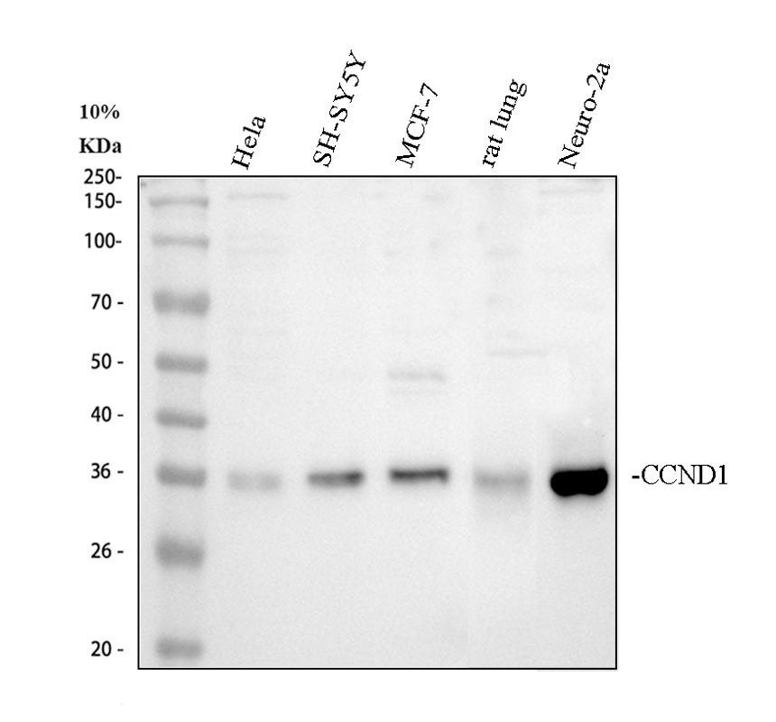
Western blot analysis of Cyclin D1/CCND1 using anti-Cyclin D1/CCND1 antibody. The sample well of each lane was loaded with 30 ug of sample under reducing conditions. After electrophoresis, proteins were transferred to a membrane. Then the membrane was incubated with rabbit anti-Cyclin D1/CCND1 antigen affinity purified polyclonal antibody at a dilution of 1:1000 and probed with a goat anti-rabbit IgG-HRP secondary antibody. The signal is developed using ECL Plus Western Blotting Substrate.IHC
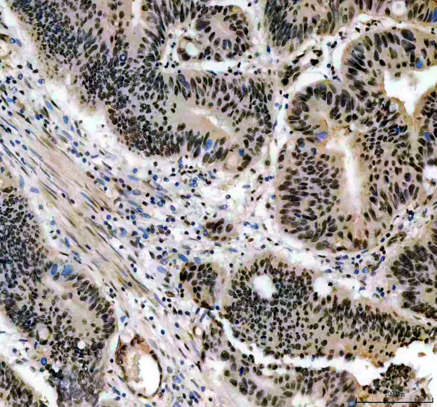
IHC analysis of Cyclin D1/CCND1 using anti-Cyclin D1/CCND1 antibody. Cyclin D1/CCND1 was detected in a paraffin-embedded section of human colon cancer tissue. The tissue section was incubated with rabbit anti-Cyclin D1/CCND1 Antibody at a dilution of 1:200 and developed using HRP Conjugated Rabbit IgG Super Vision Assay Kit with DAB as the chromogen.IHC
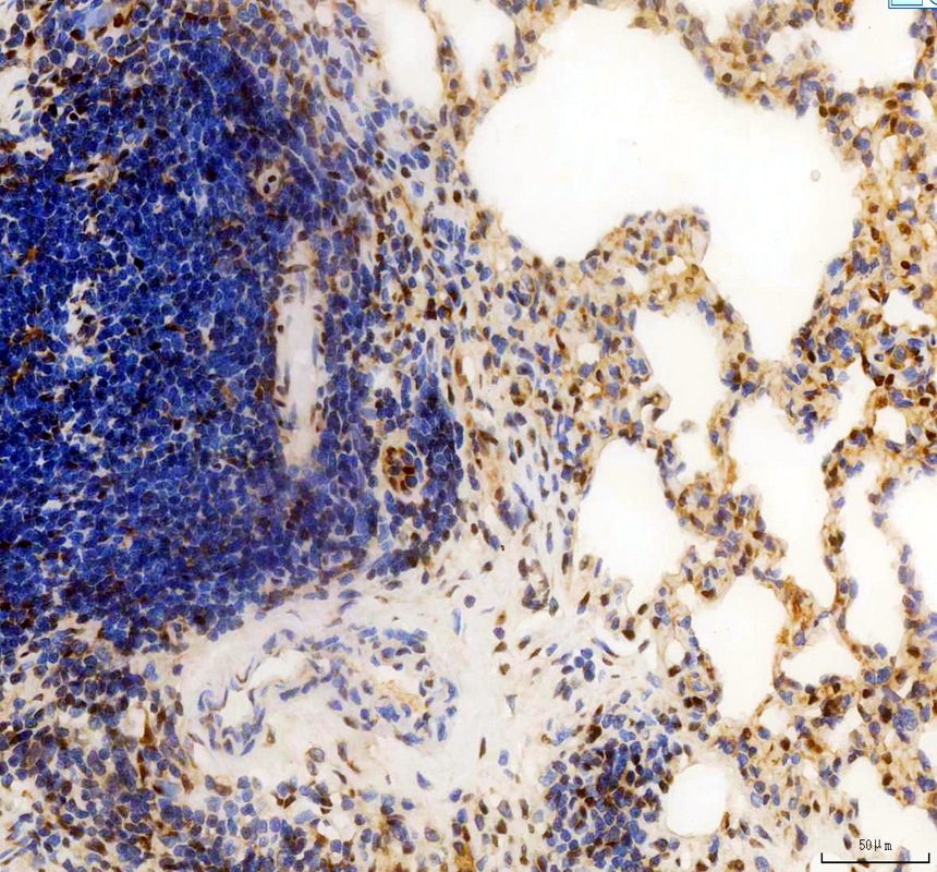
IHC analysis of Cyclin D1/CCND1 using anti-Cyclin D1/CCND1 antibody. Cyclin D1/CCND1 was detected in a paraffin-embedded section of rat lung tissue. The tissue section was incubated with rabbit anti-Cyclin D1/CCND1 Antibody at a dilution of 1:200 and developed using HRP Conjugated Rabbit IgG Super Vision Assay Kit with DAB as the chromogen.ICC/IF
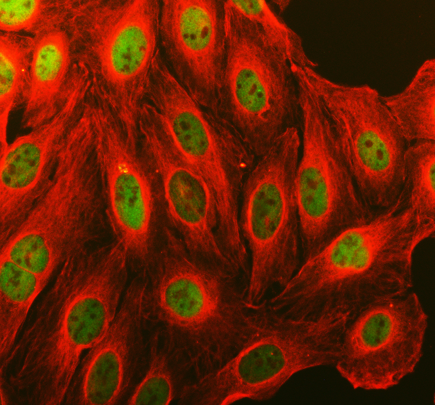
ICC/IF analysis of Cyclin D1/CCND1 using anti-Cyclin D1/CCND1 antibody and anti-Beta Tubulin antibody . Cyclin D1/CCND1 was detected in an immunocytochemical section of U2OS cells. The section was incubated with rabbit anti-Cyclin D1/CCND1 Antibody at a dilution of 1:100. Fluoro488-conjugated Anti-rabbit IgG Secondary Antibody (green) and Cy3-conjugated Anti-mouse IgG Secondary Antibody (red) were used as secondary antibody.| Product Name | Rabbit polyclonal antibody to Cyclin D1/CCND1 |
|---|---|
| Antibody Type | Primary Antibodies |
| Immunogen | A synthetic peptide corresponding to a sequence in the middle region of human Cyclin D1 different from the related mouse sequence by two amino acids, and from the related rat sequence by one amino acid. |
| Clonality | Polyclonal |
|---|---|
| Isotype | IgG |
| Host Species | Rabbit |
| Tested Applications | ICC/IFIHCWB |
| WB:1:500-1:2000 IHC:1:50-400 ICC/IF:1:50-400 |
|
| Species Reactivity | HumanMouseRat |
| Concentration | 0.5mg/ml |
| Purification | Affinity purified |
| Gene Symbol | CCND1 |
|---|---|
| Gene Synonyms | BCL1 PRAD1 U21B31 D11S287E |
| Gene Full Name | cyclin D1 |
| Gene Summary | The protein encoded by this gene belongs to the highly conserved cyclin family, whose members are characterized by a dramatic periodicity in protein abundance throughout the cell cycle. Cyclins function as regulators of CDK kinases. Different cyclins exhibit distinct expression and degradation patterns which contribute to the temporal coordination of each mitotic event. This cyclin forms a complex with and functions as a regulatory subunit of CDK4 or CDK6, whose activity is required for cell cycle G1/S transition. This protein has been shown to interact with tumor suppressor protein Rb and the expression of this gene is regulated positively by Rb. Mutations, amplification and overexpression of this gene, which alters cell cycle progression, are observed frequently in a variety of human cancers. [provided by RefSeq, Dec 2019] |
| Alternative Names | B cell lymphoma 1 protein CCND1 cyclin D Cyclin D1 D11S287E G1/S specific cyclin D1 PRAD1 PRAD1 oncogene U21B31 |
| Molecular Weight(MW) | 34 kDa |
| Cellular Localization | Cytoplasm,Membrane,Nucleus. |
WB
Western blot analysis of Cyclin D1/CCND1 using anti-Cyclin D1/CCND1 antibody. The sample well of each lane was loaded with 30 ug of sample under reducing conditions. After electrophoresis, proteins were transferred to a membrane. Then the membrane was incubated with rabbit anti-Cyclin D1/CCND1 antigen affinity purified polyclonal antibody at a dilution of 1:1000 and probed with a goat anti-rabbit IgG-HRP secondary antibody. The signal is developed using ECL Plus Western Blotting Substrate.
IHC
IHC analysis of Cyclin D1/CCND1 using anti-Cyclin D1/CCND1 antibody. Cyclin D1/CCND1 was detected in a paraffin-embedded section of human colon cancer tissue. The tissue section was incubated with rabbit anti-Cyclin D1/CCND1 Antibody at a dilution of 1:200 and developed using HRP Conjugated Rabbit IgG Super Vision Assay Kit with DAB as the chromogen.
IHC
IHC analysis of Cyclin D1/CCND1 using anti-Cyclin D1/CCND1 antibody. Cyclin D1/CCND1 was detected in a paraffin-embedded section of rat lung tissue. The tissue section was incubated with rabbit anti-Cyclin D1/CCND1 Antibody at a dilution of 1:200 and developed using HRP Conjugated Rabbit IgG Super Vision Assay Kit with DAB as the chromogen.
ICC/IF
ICC/IF analysis of Cyclin D1/CCND1 using anti-Cyclin D1/CCND1 antibody and anti-Beta Tubulin antibody . Cyclin D1/CCND1 was detected in an immunocytochemical section of U2OS cells. The section was incubated with rabbit anti-Cyclin D1/CCND1 Antibody at a dilution of 1:100. Fluoro488-conjugated Anti-rabbit IgG Secondary Antibody (green) and Cy3-conjugated Anti-mouse IgG Secondary Antibody (red) were used as secondary antibody.| Application Notes | WB:1:500-1:2000 IHC:1:50-400 ICC/IF:1:50-400 |
|---|
| Form | Liquid |
|---|---|
| Storage Instructions | 12 months from date of receipt,-20℃ as supplied. |
| Storage Buffer | 500 ug/ml antibody with PBS, 0.02% NaN3, 1 mg/ml BSA and 50% glycerol. |


