Application
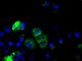
Anti-CYP1A2 mouse monoclonal antibody immunofluorescent staining of COS7 cells transiently transfected with CYP1A2Application
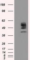
HEK293T cells were transfected with the pCMV6-ENTRY control (Left lane) or pCMV6-ENTRY CYP1A2 (Right lane) cDNA for 48 hrs and lysed. Equivalent amounts of cell lysates (5 ug per lane) were separated by SDS-PAGE and immunoblotted with anti-CYP1A2.Application
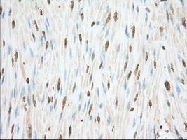
Immunohistochemical staining of paraffin-embedded Human colon tissue using anti-CYP1A2 mouse monoclonal antibody. ( Dilution 1:50)Application
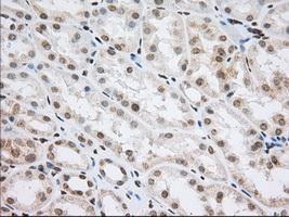
Immunohistochemical staining of paraffin-embedded Human Kidney tissue using anti-CYP1A2 mouse monoclonal antibody. ( Dilution 1:50)Application
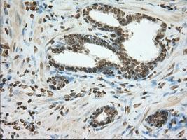
Immunohistochemical staining of paraffin-embedded Carcinoma of Human prostate tissue using anti-CYP1A2 mouse monoclonal antibody. ( Dilution 1:50)Application
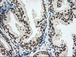
Immunohistochemical staining of paraffin-embedded Human prostate tissue using anti-CYP1A2 mouse monoclonal antibody. ( Dilution 1:50)Application
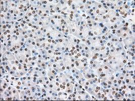
Immunohistochemical staining of paraffin-embedded Human pancreas tissue using anti-CYP1A2 mouse monoclonal antibody. ( Dilution 1:50)Application
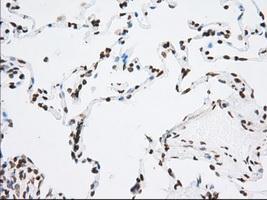
Immunohistochemical staining of paraffin-embedded Human lung tissue using anti-CYP1A2 mouse monoclonal antibody. ( Dilution 1:50)Application
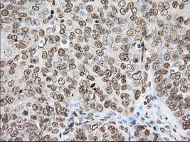
Immunohistochemical staining of paraffin-embedded Adenocarcinoma of Human ovary tissue using anti-CYP1A2 mouse monoclonal antibody. ( Dilution 1:50)Application
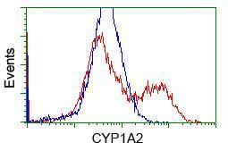
HEK293T cells transfected with either RC221636 overexpress plasmid(Red) or empty vector control plasmid(Blue) were immunostained by anti-CYP1A2 antibody, and then analyzed by flow cytometry.Application
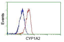
Flow cytometric Analysis of Jurkat cells, using anti-CYP1A2 antibody,(Red), compared to a nonspecific negative control antibody(TA50011),(Blue).Application
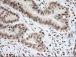
Immunohistochemical staining of paraffin-embedded Adenocarcinoma of Human colon tissue using anti-CYP1A2 mouse monoclonal antibody. ( Dilution 1:50)| Product Name | CYP1A2 antibody |
|---|---|
| Antibody Type | Primary Antibodies |
| Product description | Mouse monoclonal antibody [8F1] to CYP1A2Cytochromes P450 are a group of heme-thiolate monooxygenases. In liver microsomes, this enzyme is involved in an NADPH-dependent electron transport pathway. It oxidizes a variety of structurally unrelated compounds, including steroids, fatty acids, and xenobiotics. Most active in catalyzing 2-hydroxylation. Caffeine is metabolized primarily by cytochrome CYP1A2 in the liver through an initial N3-demethylation. Also acts in the metabolism of aflatoxin B1 and acetaminophen. Participates in the bioactivation of carcinogenic aromatic and heterocyclic amines. Catalizes the N-hydroxylation of heterocyclic amines and the O-deethylation of phenacetin. |
| Immunogen | Full length human recombinant protein of human CYP1A2 (NP_000752) produced in HEK293T cell. |
| Clonality | Monoclonal |
|---|---|
| Isotype | IgG2a |
| Clone Number | 8F1 |
| Host Species | Mouse |
| Tested Applications | FACSICC/IFIHCIHC-PWB |
| IHC 1:50 : |
|
| Species Reactivity | Human |
| Concentration | 1mg/ml |
| Purification | Affinity purified |
| Gene Synonyms | CYP1A2 CP12 P450(PA) P3450 P3-450 |
|---|---|
| Target | CYP1A2 |
| Tissue Specificity | is specific for human CYP1A2 (NP_000752). |
Application
Anti-CYP1A2 mouse monoclonal antibody immunofluorescent staining of COS7 cells transiently transfected with CYP1A2
Application
HEK293T cells were transfected with the pCMV6-ENTRY control (Left lane) or pCMV6-ENTRY CYP1A2 (Right lane) cDNA for 48 hrs and lysed. Equivalent amounts of cell lysates (5 ug per lane) were separated by SDS-PAGE and immunoblotted with anti-CYP1A2.
Application
Immunohistochemical staining of paraffin-embedded Human colon tissue using anti-CYP1A2 mouse monoclonal antibody. ( Dilution 1:50)
Application
Immunohistochemical staining of paraffin-embedded Human Kidney tissue using anti-CYP1A2 mouse monoclonal antibody. ( Dilution 1:50)
Application
Immunohistochemical staining of paraffin-embedded Carcinoma of Human prostate tissue using anti-CYP1A2 mouse monoclonal antibody. ( Dilution 1:50)
Application
Immunohistochemical staining of paraffin-embedded Human prostate tissue using anti-CYP1A2 mouse monoclonal antibody. ( Dilution 1:50)
Application
Immunohistochemical staining of paraffin-embedded Human pancreas tissue using anti-CYP1A2 mouse monoclonal antibody. ( Dilution 1:50)
Application
Immunohistochemical staining of paraffin-embedded Human lung tissue using anti-CYP1A2 mouse monoclonal antibody. ( Dilution 1:50)
Application
Immunohistochemical staining of paraffin-embedded Adenocarcinoma of Human ovary tissue using anti-CYP1A2 mouse monoclonal antibody. ( Dilution 1:50)
Application
HEK293T cells transfected with either RC221636 overexpress plasmid(Red) or empty vector control plasmid(Blue) were immunostained by anti-CYP1A2 antibody, and then analyzed by flow cytometry.
Application
Flow cytometric Analysis of Jurkat cells, using anti-CYP1A2 antibody,(Red), compared to a nonspecific negative control antibody(TA50011),(Blue).
Application
Immunohistochemical staining of paraffin-embedded Adenocarcinoma of Human colon tissue using anti-CYP1A2 mouse monoclonal antibody. ( Dilution 1:50)| Application Notes | IHC 1:50 : |
|---|
| Form | Liquid |
|---|---|
| Storage Instructions | For short-term storage, store at 4°C or aliquot into working amounts and store at -20°C. For long-term storage, store at -70°C (aliquotted). Avoid repeated freeze-thaw cycles. |
| Storage Buffer | PBS (PH 7.3) containing 1% BSA, 50% glycerol and 0.02% sodium azide. |


