Application
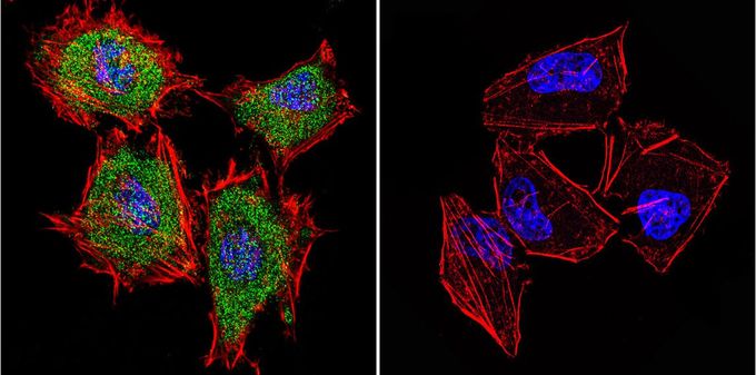
Immunofluorescent analysis of Cytohesin 1 in HeLa Cells. Cells were grown on slides and fixed with formaldehyde prior to staining. Cells were probed without (control) or with Cytohesin 1 antibody [2E11] at a dilution of 1:20 overnight at 4 C, washed with PBS and incubated with a proper secondary antibody. Cytohesin 1 staining (green), F-Actin staining with Phalloidin (red) and nuclei with DAPI (blue) is shown. Images were taken at 60X magnification.| Product Name | Cytohesin 1 antibody |
|---|---|
| Antibody Type | Primary Antibodies |
| Product description | Mouse monoclonal antibody [2E11] to Cytohesin 1The protein encoded by this gene is a member of the PSCD family. Members of this family have identical structural organization that consists of an N-terminal coiled-coil motif, a central Sec7 domain, and a C-terminal pleckstrin homology (PH) domain. The coiled-coil motif is involved in homodimerization, the Sec7 domain contains guanine-nucleotide exchange protein (GEP) activity, and the PH domain interacts with phospholipids and is responsible for association of PSCDs with membranes. Members of this family appear to mediate the regulation of protein sorting and membrane trafficking. This gene is highly expressed in natural killer and peripheral T cells, and regulates the adhesiveness of integrins at the plasma membrane of lymphocytes. The encoded protein is 83% homologous to that of CYTH2. [provided by RefSeq, Aug 2008] |
| Immunogen | Purified, His-tagged cytohesin-1. |
| Clonality | Monoclonal |
|---|---|
| Isotype | IgG1 |
| Clone Number | 2E11 |
| Host Species | Mouse |
| Tested Applications | ICC/IFIPWB |
| ICC/IF: 1:20: |
|
| Species Reactivity | HumanMouse |
| Concentration | 1mg/ml (Please refer to the vial label for the specific concentration) |
| Purification | Affinity purified |
| Gene Synonyms | cytohesin 1 D17S811E PSCD1 CYTOHESIN-1 B2-1 SEC7 CYTH1 |
|---|---|
| Target | Cytohesin 1 |
Application
Immunofluorescent analysis of Cytohesin 1 in HeLa Cells. Cells were grown on slides and fixed with formaldehyde prior to staining. Cells were probed without (control) or with Cytohesin 1 antibody [2E11] at a dilution of 1:20 overnight at 4 C, washed with PBS and incubated with a proper secondary antibody. Cytohesin 1 staining (green), F-Actin staining with Phalloidin (red) and nuclei with DAPI (blue) is shown. Images were taken at 60X magnification.| Application Notes | ICC/IF: 1:20: |
|---|
| Form | Liquid |
|---|---|
| Storage Instructions | -20° C, Avoid Freeze/Thaw Cycles |
| Storage Buffer | PBS containing 0.05 % sodium azide and 1 mg/ml BSA |


