Application
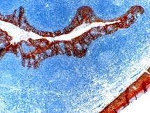
IHC Image using - Detection of Cytokeratin by IHC in Human TonsilApplication
Formalin fixed paraffin embedded human tonsil stained with Cytokeratin antibody .| Product Name | Cytokeratin antibody |
|---|---|
| Antibody Type | Primary Antibodies |
| Product description | Mouse monoclonal [MNF116] to CytokeratinCytokeratins, a group of at least 29 different proteins, are characteristic of epithelial and trichocytic cells. Cytokeratins 4, 5, 6 and 8 are members of the type II neutral-to-basic subfamily. Cytokeratin peptide 4 (59 kDa) is the secondary type II keratin expressed in non cornified stratified squamous epithelia. Cytokeratin peptide 5 (58 kDa) is the primary type II keratin in stratified epithelia, while cytokeratin type 8 (52 kDa) is a major type II keratin in simple epithelia. Cytokeratin 6 (56 kDa) is a "hyperproliferation" cytokeratin expressed in tissues with natural or pathological high turnover. Cytokeratins 10, 13 and 18 are members of the type I acidic subfamily. Cytokeratin peptide 10 (56 kDa) is the secondary type I keratin expressed in cornified epithelia. Cytokeratin 13 (54 kDa) is the secondary type I keratin expressed in non-cornified stratified squamous epithelia. Cytokeratin 18 (45 kDa) is the primary type I keratin expressed in simple epithelial cells.Cytokeratin is a heterotetramer of two type I and two type II keratins. Keratin 1 is generally associated with keratin 10. Defects in Cytokeratin are a cause of epidermolytic hyperkeratosis, also known as bullous congenital ichthyosiform erythroderma (BIE), which is a hereditary skin disorder characterized by blistering and a marked thickening of the stratum corneum. Defects in Cytokeratin are also the cause of Curth Macklin type ichthyosis hystrix, nonepidermolytic palmoplantar keratoderma, annular epidermolytic ichthyosis and keratosis palmoplantaris striata III (PPKS3). PPKS3 is an autosomal dominant disorder affecting palm and sole skin where stratum corneum and epidermal layers are thickened. There is no involvement of nonpalmoplantar skin, and both hair and nails are normal. |
| Immunogen | Crude extract of splenic cells from a nude mouse engrafted with MCF-7 cells. |
| Clonality | Monoclonal |
|---|---|
| Isotype | IgG1 |
| Clone Number | MNF116 |
| Host Species | Mouse |
| Tested Applications | IHCIHC-P |
| For IHC-P: Use at a dilution of 1:25-1:50 in an ABC method. Suggested incubation period of 30mins at RT. Enzymatic predigestion of paraffin embedded tissues is recommended prior to the immunostaining. Optimal dilutions/concentrations should be determined by the researcher.: |
|
| Species Reactivity | Human |
| Concentration | 1mg/ml |
| Purification | Affinity purified |
| Gene Synonyms | EHK1 P04264 3848 CK1 KRT1A 113800 KRT1 K1 Cytokeratin |
|---|---|
| Target | Cytokeratin |
| Tissue Specificity | Recognizes cytokeratin polypeptides ranging from 40kDa through 58kD, corresponding to cytokeratins 5, 6, 8, 17 and 19. Shows a broad pattern of reactivity with human epithelial tissues from simple glandular epithelia to stratified squamous epithelia, like epidermis, mammary gland ducts and tracheal epithelium. |
| Light Chain Type | kappa |
Application
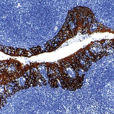
IHC Image using - Detection of Cytokeratin by IHC in Human Tonsil
Application
Formalin fixed paraffin embedded human tonsil stained with Cytokeratin antibody .| Positive Control | Skin |
|---|---|
| Application Notes | For IHC-P: Use at a dilution of 1:25-1:50 in an ABC method. Suggested incubation period of 30mins at RT. Enzymatic predigestion of paraffin embedded tissues is recommended prior to the immunostaining. Optimal dilutions/concentrations should be determined by the researcher.: |
| Form | Liquid |
|---|---|
| Storage Instructions | Keep as concentrated solution, aliquot and store at 4°C. Do not freeze. |
| Storage Buffer | Tissue culture supernatant containing sodium azide |


