Application
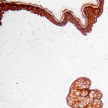
Detection of Cytokeratin high MW by IHC in Human SkinApplication
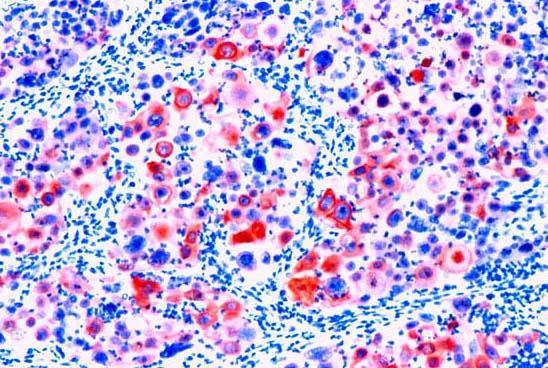
Cytokeratin H. M. Wt.Formalin fixed paraffin embedded human colon carcinoma stained with Cytokeratin, H.M.W. (cat. # ) using ABC and AEC chromogen.| Product Name | Cytokeratin high MW antibody |
|---|---|
| Antibody Type | Primary Antibodies |
| Product description | Monoclonal Mouse Antibody to Human Cytokeratin ( H.M.Weight )This antibody recognizes keratin polypeptide of 68, 58, 56.5 and 50 kD in extract of stratum corneum. The antibody reacts with squamous, ductal and other complex epithelia. It stains with adenocarcinomas, breast, pancreas, bile duct, salivary gland and transitional cell carcinomas. |
| Immunogen | BALB/C mice were immunized with solublized keratin extract from human stratum corneum. |
| Clonality | Monoclonal |
|---|---|
| Isotype | IgG1 |
| Clone Number | 34ßE12 |
| Host Species | Mouse |
| Tested Applications | IHC-P |
| This antibody may be diluted to a titer of 1:25-1:50 in an ABC method. However, depending on the staining conditions employed, we suggest that final dilution should be determined by the user. We suggest an incubation period of 30 minutes at room temperature. However, depending upon the fixation conditions and the staining system employed, optimal incubation should be determined by the user. Enzymatic predigestion of paraffin embedded tissues is recommended prior to the immunostaining.: |
|
| Species Reactivity | Human |
| Concentration | 1mg/ml |
| Gene Synonyms | EHK1 P04264 3848 Cytokeratin high MW CK1 KRT1A 113800 KRT1 K1 |
|---|---|
| Target | Cytokeratin high MW |
| Cellular Localization | Cytoplasmic |
| Light Chain Type | Kappa |
Application
Detection of Cytokeratin high MW by IHC in Human Skin
Application
Cytokeratin H. M. Wt.Formalin fixed paraffin embedded human colon carcinoma stained with Cytokeratin, H.M.W. (cat. # ) using ABC and AEC chromogen.| Positive Control | Skin |
|---|---|
| Application Notes | This antibody may be diluted to a titer of 1:25-1:50 in an ABC method. However, depending on the staining conditions employed, we suggest that final dilution should be determined by the user. We suggest an incubation period of 30 minutes at room temperature. However, depending upon the fixation conditions and the staining system employed, optimal incubation should be determined by the user. Enzymatic predigestion of paraffin embedded tissues is recommended prior to the immunostaining.: |
| Form | Liquid |
|---|---|
| Storage Instructions | Keep as concentrated solution, aliquot and store at 4°C. |
| Storage Buffer | phosphate buffered saline , pH 7.4, 150mM NaCl, 0.02% sodium azide and 50% glycerol. |


