Application
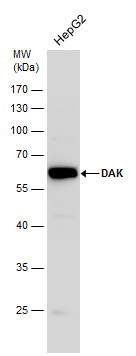
DAK antibody detects DAK protein by western blot analysis. Whole cell extracts (30 μg) was separated by 10% SDS-PAGE, and the membrane was blotted with DAK antibody diluted by 1:1000.| Product Name | DAK antibody |
|---|---|
| Antibody Type | Primary Antibodies |
| Product description | Rabbit Polyclonal antibody to DAK (dihydroxyacetone kinase 2 homolog (S. cerevisiae))This gene is a member of the family of dihydroxyacetone kinases, which have a protein structure distinct from other kinases. The product of this gene phosphorylates dihydroxyacetone, and also catalyzes the formation of riboflavin 4',5'-phosphate (aka cyclin FMN) from FAD. Several alternatively spliced transcript variants have been identified, but the full-length nature of only one has been determined. [provided by RefSeq] |
| Immunogen | Recombinant protein encompassing a sequence within the center region of human DAK. The exact sequence is proprietary. |
| Clonality | Polyclonal |
|---|---|
| Isotype | IgG |
| Host Species | Rabbit |
| Tested Applications | WB |
| *Optimal dilutions/concentrations should be determined by the researcher.
: |
|
| Species Reactivity | Human |
| Predicted Reactivity | Mouse, Rat, Chimpanzee, BovineMouse (83%), Rat (83%), Chimpanzee (100%), Bovine (89%) |
| Concentration | 1mg/ml (Please refer to the vial label for the specific concentration) |
| Purification | Affinity purified |
| Gene Synonyms | NET45 dihydroxyacetone kinase 2 homolog (S. cerevisiae) |
|---|---|
| Target | DAK |
| Molecular Weight(MW) | 59 kDa (note) |
Application
DAK antibody detects DAK protein by western blot analysis. Whole cell extracts (30 μg) was separated by 10% SDS-PAGE, and the membrane was blotted with DAK antibody diluted by 1:1000.| Positive Control | HepG2 |
|---|---|
| Application Notes | *Optimal dilutions/concentrations should be determined by the researcher.
: |
| Form | Liquid |
|---|---|
| Storage Instructions | Keep as concentrated solution. Aliquot and store at -20ºC or below. Avoid multiple freeze-thaw cycles. |
| Storage Buffer | 1XPBS, 20% Glycerol (pH7). 0.01% Thimerosal was added as a preservative. |


