Application
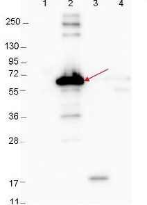
Western blot showing detection of 0.1 µg recombinant proteins in western blot. Lane 1: Molecular weight markers. Lane 2: MBP-DbpA fusion protein (arrow; expected MW: 60.9 kDa). Lane 3: DbpA, MBP removed by TEV cleavage. Lane 4: MBP alone. Protein was run on a 4-20% gel, then transferred to 0.45 µm nitrocellulose. After blocking with 1% BSA-TTBS overnight at 4°C, primary antibody was used at 1:1000 at room temperature for 30 min. HRP-conjugated Goat-Anti-Rabbit secondary antibody was used at 1:40,000 in blocking buffer and imaged on the VersaDoc MP 4000 imaging system (Bio-Rad).| Product Name | Dbp-A antibody |
|---|---|
| Antibody Type | Primary Antibodies |
| Product description | Rabbit Polyclonal antibody to Dbp-AThis product is antibody made against DbpA, or Decorin Binding Protein A from the spirochete Borrelia burgdorferi, which is carried by Ixodes ticks. DbpA from other microbial organisms such as E. coli (ATP-dependent RNA helicase DbpA) are significantly different. The spirochete migrates from the tick midgut during tick feeding to tick salivary glands and are thus transmitted to the mammal host. This transition may be facilitated by changes in expression of some B. burgdorferi genes. Spirochetal surface adhesions mediate attachment to decorin, a major component of the host extracellular matrix, enabling bacteria to colonize in mammalian tissues. It is believed that expression of the various proteins associated with the spirochete may be regulated by the changes in tick life cycle, changes in conditions during tick feeding (such as temperature, pH, and nutrients) and/or in coordination with the course of infection of the mammal host. |
| Immunogen | MBP-fusion protein corresponding to Borrelia burgdorferi Dbp-A protein. |
| Clonality | Polyclonal |
|---|---|
| Isotype | IgG |
| Host Species | Rabbit |
| Tested Applications | ELISAWB |
| *Optimal dilutions/concentrations should be determined by the researcher.
: |
|
| Concentration | 1mg/ml (Please refer to the vial label for the specific concentration) |
| Purification | Protein A |
| Gene Synonyms | DBPA |
|---|---|
| Target | Dbp-A |
Application
Western blot showing detection of 0.1 µg recombinant proteins in western blot. Lane 1: Molecular weight markers. Lane 2: MBP-DbpA fusion protein (arrow; expected MW: 60.9 kDa). Lane 3: DbpA, MBP removed by TEV cleavage. Lane 4: MBP alone. Protein was run on a 4-20% gel, then transferred to 0.45 µm nitrocellulose. After blocking with 1% BSA-TTBS overnight at 4°C, primary antibody was used at 1:1000 at room temperature for 30 min. HRP-conjugated Goat-Anti-Rabbit secondary antibody was used at 1:40,000 in blocking buffer and imaged on the VersaDoc MP 4000 imaging system (Bio-Rad).| Application Notes | *Optimal dilutions/concentrations should be determined by the researcher.
: |
|---|
| Form | Liquid |
|---|---|
| Storage Instructions | Store vial at 4° C for short-term use.For extended storage aliquot contents and freeze at -20° C or below. Avoid cycles of freezing and thawing. |
| Storage Buffer | 0.02 M Potassium Phosphate, 0.15 M Sodium Chloride, pH 7.2 and 0.01% (w/v) Sodium Azide |


