Application
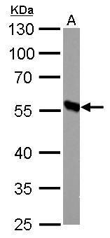
DBT antibody detects DBT protein by western blot analysis. A. 50 µg mouse liver lysate/extract 10% SDS-PAGE DBT antibody dilution: 1:1000 The HRP-conjugated anti-rabbit IgG antibody was used to detect the primary antibody.Application
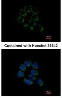
Immunofluorescence analysis of methanol-fixed A431, using DBT antibody at 1:200 dilution.Application
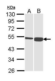
Sample (30 µg of whole cell lysate) A: H1299 B: HepG2 10% SDS PAGE diluted at 1:1000 The HRP-conjugated anti-rabbit IgG antibody was used to detect the primary antibody.Application
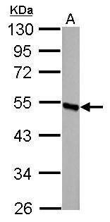
DBT antibody detects DBT protein by western blot analysis. A. 50 µg rat liver lysate/extract 10% SDS-PAGE DBT antibody dilution: 1:1000 The HRP-conjugated anti-rabbit IgG antibody was used to detect the primary antibody.Application
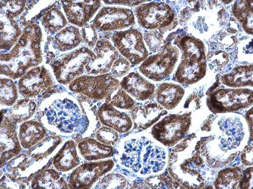
DBT antibody detects DBT protein at mitochondria on mouse kidney by immunohistochemical analysis. Sample: Paraffin-embedded mouse kidney. DBT antibody diluted at 1:500.| Product Name | DBT antibody |
|---|---|
| Antibody Type | Primary Antibodies |
| Product description | Rabbit Polyclonal antibody to DBT (dihydrolipoamide branched chain transacylase E2)The branched-chain alpha-keto acid dehydrogenase complex (BCKD) is an inner-mitochondrial enzyme complex involved in the breakdown of the branched-chain amino acids isoleucine, leucine, and valine. The BCKD complex is thought to be composed of a core of 24 transacylase (E2) subunits, and associated decarboxylase (E1), dehydrogenase (E3), and regulatory subunits. This gene encodes the transacylase (E2) subunit. Mutations in this gene result in maple syrup urine disease, type 2. Alternatively spliced transcript variants have been described, but their biological validity has not been determined. [provided by RefSeq] |
| Immunogen | Recombinant protein encompassing a sequence within the center region of human DBT. The exact sequence is proprietary. |
| Clonality | Polyclonal |
|---|---|
| Isotype | IgG |
| Host Species | Rabbit |
| Tested Applications | ICC/IFIHC-PWB |
| *Optimal dilutions/concentrations should be determined by the researcher.
: |
|
| Species Reactivity | HumanMouseRat |
| Predicted Reactivity | Zebrafish, Xenopus laevis, Chicken, Rhesus Monkey, Chimpanzee, BovineZebrafish (82%), Xenopus laevis (89%), Chicken (85%), Rhesus Monkey (100%), Chimpanzee (100%), Bovine (96%) |
| Concentration | 1mg/ml |
| Purification | Affinity purified |
| Gene Synonyms | BCATE2 antibody E2 antibody E2B antibody MGC9061 antibody DBT antibody BCKAD E2 subunit antibody dihydrolipoyllysine-residue (2-methylpropanoyl)transferase antibody dihydrolipoyl transacylase antibody mitochondrial branched chain alpha-keto acid de |
|---|---|
| Molecular Weight(MW) | 53 kDa (note) (2) |
| Cellular Localization | Mitochondrion matrix |
Application
DBT antibody detects DBT protein by western blot analysis. A. 50 µg mouse liver lysate/extract 10% SDS-PAGE DBT antibody dilution: 1:1000 The HRP-conjugated anti-rabbit IgG antibody was used to detect the primary antibody.
Application
Immunofluorescence analysis of methanol-fixed A431, using DBT antibody at 1:200 dilution.
Application
Sample (30 µg of whole cell lysate) A: H1299 B: HepG2 10% SDS PAGE diluted at 1:1000 The HRP-conjugated anti-rabbit IgG antibody was used to detect the primary antibody.
Application
DBT antibody detects DBT protein by western blot analysis. A. 50 µg rat liver lysate/extract 10% SDS-PAGE DBT antibody dilution: 1:1000 The HRP-conjugated anti-rabbit IgG antibody was used to detect the primary antibody.
Application
DBT antibody detects DBT protein at mitochondria on mouse kidney by immunohistochemical analysis. Sample: Paraffin-embedded mouse kidney. DBT antibody diluted at 1:500.| Positive Control | H1299 , HepG2 , mouse liver , rat liver |
|---|---|
| Application Notes | *Optimal dilutions/concentrations should be determined by the researcher.
: |
| Form | Liquid |
|---|---|
| Storage Instructions | Keep as concentrated solution. Aliquot and store at -20ºC or below. Avoid multiple freeze-thaw cycles. |
| Storage Buffer | 0.1M Tris, 0.1M Glycine, 10% Glycerol (pH7). 0.01% Thimerosal was added as a preservative. |


