Application
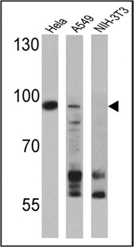
Western blot analysis of DNA Methyltransferase 3 was performed by loading 25 ug of Hela (Lane 1), A549 (Lane 2), and NIH-3T3 (Lane 3) cell lysates and a molecular weight protein ladder onto an SDS polyacrylamide gel. Proteins were transferred to a PVDF membrane and blocked with a blocking buffer at 4ºC overnight. The membrane was probed with a DNA Methyltransferase 3 polyclonal antibody at a dilution of 1:1000 overnight at 4°C, washed in TBST, and probed with an HRP-conjugated secondary antibody for 1 hr at room temperature in the dark. Results show a band at 97 kDa in A549 and Hela cell lysates.Application
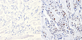
Immunohistochemistry analysis of DNA Methyltransferase 3 showing positive staining in the nucleus and cytoplasm of paraffin-treated Human breast carcinoma (right) compared with a negative control in the absence of primary antibody (left). To expose target proteins, antigen retrieval method was performed using 10mM sodium citrate (pH 6.0) microwaved for 8-15 min. Following antigen retrieval, tissues were blocked in 3% H2O2-methanol for 15 min at room temperature, washed with ddH2O and PBS, and then probed with a DNA Methyltransferase 3 polyclonal antibody diluted by 3% BSA-PBS at a dilution of 1:500 overnight at 4°C in a humidified chamber. Tissues were washed extensively PBST and detection was performed using an HRP-conjugated secondary antibody followed by colorimetric detection using a DAB kit. Tissues were counterstained with hematoxylin and dehydrated with ethanol and xylene to prep for mounting.Application
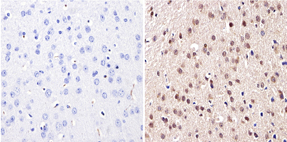
Immunohistochemistry analysis of DNA Methyltransferase 3 showing positive staining in the nucleus and cytoplasm of paraffin-treated Mouse brain tissue (right) compared with a negative control in the absence of primary antibody (left). To expose target proteins, antigen retrieval method was performed using 10mM sodium citrate (pH 6.0) microwaved for 8-15 min. Following antigen retrieval, tissues were blocked in 3% H2O2-methanol for 15 min at room temperature, washed with ddH2O and PBS, and then probed with a DNA Methyltransferase 3 polyclonal antibody diluted by 3% BSA-PBS at a dilution of 1:200 overnight at 4°C in a humidified chamber. Tissues were washed extensively PBST and detection was performed using an HRP-conjugated secondary antibody followed by colorimetric detection using a DAB kit. Tissues were counterstained with hematoxylin and dehydrated with ethanol and xylene to prep for mounting.Application
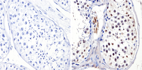
Immunohistochemistry analysis of DNA Methyltransferase 3 showing positive staining in the nucleus of paraffin-treated Human testis tissue (right) compared with a negative control in the absence of primary antibody (left). To expose target proteins, antigen retrieval method was performed using 10mM sodium citrate (pH 6.0) microwaved for 8-15 min. Following antigen retrieval, tissues were blocked in 3% H2O2-methanol for 15 min at room temperature, washed with ddH2O and PBS, and then probed with a DNA Methyltransferase 3 polyclonal antibody diluted by 3% BSA-PBS at a dilution of 1:500 overnight at 4°C in a humidified chamber. Tissues were washed extensively PBST and detection was performed using an HRP-conjugated secondary antibody followed by colorimetric detection using a DAB kit. Tissues were counterstained with hematoxylin and dehydrated with ethanol and xylene to prep for mounting.| Product Name | DNMT3B antibody |
|---|---|
| Antibody Type | Primary Antibodies |
| Product description | Rabbit polyclonal antibody to DNMT3BDNA methyltransferases methylate the 5-position of cytosine in the context of CpG dinucleotides. DNA methylation is crucial for normal embryonic development, genetic imprinting, and X-chromosome inactivation. Hypermethylation of DNA has also been implicated in tumorigenesis through improper methylation of growth regulatory gene promoter regions. |
| Immunogen | Synthetic peptide corresponding to the residues M(1) K G D S R H L N E E E G A(14) C of mouse DNMT3b |
| Clonality | Polyclonal |
|---|---|
| Host Species | Rabbit |
| Tested Applications | IHC-FIHC-PWB |
| IHC-P: 1:100 - 1:1000: |
|
| Species Reactivity | HumanMouse |
| Concentration | 1mg/ml (Please refer to the vial label for the specific concentration) |
| Purification | Affinity purified |
| Gene Synonyms | ICF DNMT3B M.HsaIIIB MGC124407 242860 1789 13436 Q9UBC3 |
|---|---|
| Target | DNA Methyltransferase 3 |
Application
Western blot analysis of DNA Methyltransferase 3 was performed by loading 25 ug of Hela (Lane 1), A549 (Lane 2), and NIH-3T3 (Lane 3) cell lysates and a molecular weight protein ladder onto an SDS polyacrylamide gel. Proteins were transferred to a PVDF membrane and blocked with a blocking buffer at 4ºC overnight. The membrane was probed with a DNA Methyltransferase 3 polyclonal antibody at a dilution of 1:1000 overnight at 4°C, washed in TBST, and probed with an HRP-conjugated secondary antibody for 1 hr at room temperature in the dark. Results show a band at 97 kDa in A549 and Hela cell lysates.
Application
Immunohistochemistry analysis of DNA Methyltransferase 3 showing positive staining in the nucleus and cytoplasm of paraffin-treated Human breast carcinoma (right) compared with a negative control in the absence of primary antibody (left). To expose target proteins, antigen retrieval method was performed using 10mM sodium citrate (pH 6.0) microwaved for 8-15 min. Following antigen retrieval, tissues were blocked in 3% H2O2-methanol for 15 min at room temperature, washed with ddH2O and PBS, and then probed with a DNA Methyltransferase 3 polyclonal antibody diluted by 3% BSA-PBS at a dilution of 1:500 overnight at 4°C in a humidified chamber. Tissues were washed extensively PBST and detection was performed using an HRP-conjugated secondary antibody followed by colorimetric detection using a DAB kit. Tissues were counterstained with hematoxylin and dehydrated with ethanol and xylene to prep for mounting.
Application
Immunohistochemistry analysis of DNA Methyltransferase 3 showing positive staining in the nucleus and cytoplasm of paraffin-treated Mouse brain tissue (right) compared with a negative control in the absence of primary antibody (left). To expose target proteins, antigen retrieval method was performed using 10mM sodium citrate (pH 6.0) microwaved for 8-15 min. Following antigen retrieval, tissues were blocked in 3% H2O2-methanol for 15 min at room temperature, washed with ddH2O and PBS, and then probed with a DNA Methyltransferase 3 polyclonal antibody diluted by 3% BSA-PBS at a dilution of 1:200 overnight at 4°C in a humidified chamber. Tissues were washed extensively PBST and detection was performed using an HRP-conjugated secondary antibody followed by colorimetric detection using a DAB kit. Tissues were counterstained with hematoxylin and dehydrated with ethanol and xylene to prep for mounting.
Application
Immunohistochemistry analysis of DNA Methyltransferase 3 showing positive staining in the nucleus of paraffin-treated Human testis tissue (right) compared with a negative control in the absence of primary antibody (left). To expose target proteins, antigen retrieval method was performed using 10mM sodium citrate (pH 6.0) microwaved for 8-15 min. Following antigen retrieval, tissues were blocked in 3% H2O2-methanol for 15 min at room temperature, washed with ddH2O and PBS, and then probed with a DNA Methyltransferase 3 polyclonal antibody diluted by 3% BSA-PBS at a dilution of 1:500 overnight at 4°C in a humidified chamber. Tissues were washed extensively PBST and detection was performed using an HRP-conjugated secondary antibody followed by colorimetric detection using a DAB kit. Tissues were counterstained with hematoxylin and dehydrated with ethanol and xylene to prep for mounting.| Application Notes | IHC-P: 1:100 - 1:1000: |
|---|
| Form | Liquid |
|---|---|
| Storage Instructions | Keep as concentrated solution. Aliquot and store at -20ºC or below. Avoid multiple freeze-thaw cycles. |
| Storage Buffer | PBS containing 1 mg/ml BSA and 0.05% sodium azide. |


