Application
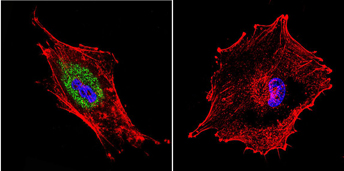
Immunofluorescent analysis of Dynein in U87-MG Cells. Cells were grown on chamber slides and fixed with formaldehyde prior to staining. Cells were probed without (control) or with a Dynein monoclonal antibody at a dilution of 1:20 overnight at 4 C, washed with PBS and incubated with a DyLight-488 conjugated secondary antibody. Dynein staining (green), F-Actin staining with Phalloidin (red) and nuclei with DAPI (blue) is shown.Application
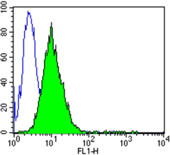
Flow cytometry analysis of Dynein showing positive staining in the cytoplasm of C6 cells compared to an isotype control (blue). Cells were harvested, adjusted to a concentration of 1-5x10^6 cells/ml, fixed with 2% paraformaldehyde and washed with PBS. Cells were penetrated by dropping the supernatant, adding 90% methanol and incubated for 10 minutes at room temperature. Follwing penetration, cells were blocked with a 2% solution of BSA-PBS for 30 min at room temperature and incubated with a Dynein monoclonal antibody at a dilution of 1 ug/test for 60 min at room temperature. Cells were then incubated for 40 min at room temperature in the dark using a Dylight 488-conjugated goat anti-mouse IgG (H+L) secondary antibody and re-suspended in PBS for FACS analysis.Application
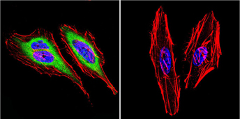
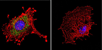
Immunofluorescent analysis of Dynein in HeLa Cells. Cells were grown on chamber slides and fixed with formaldehyde prior to staining. Cells were probed without (control) or with a Dynein monoclonal antibody at a dilution of 1:20 overnight at 4 C, washed with PBS and incubated with a DyLight-488 conjugated secondary antibody. Dynein staining (green), F-Actin staining with Phalloidin (red) and nuclei with DAPI (blue) is shown.Application
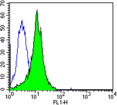
Flow cytometry analysis of Dynein showing positive staining in the cytoplasm of U87-MG cells compared to an isotype control (blue). Cells were harvested, adjusted to a concentration of 1-5x10^6 cells/ml, fixed with 2% paraformaldehyde and washed with PBS. Cells were penetrated by dropping the supernatant, adding 90% methanol and incubated for 10 minutes at room temperature. Following penetration, cells were blocked with a 2% solution of BSA-PBS for 30 min at room temperature and incubated with a Dynein monoclonal antibody at a dilution of 2 ug/test for 60 min at room temperature. Cells were then incubated for 40 min at room temperature in the dark using a Dylight 488-conjugated goat anti-mouse IgG (H+L) secondary antibody and re-suspended in PBS for FACS analysis.Application
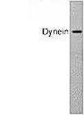
Western blot of cytoplasmic dynein in HeLa cell lysate usingApplication
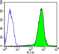
Flow cytometry analysis of Dynein showing strong positive staining in the cytoplasm of Hela cells compared to an isotype control (blue). Cells were harvested, adjusted to a concentration of 1-5x10^6 cells/ml, fixed with 2% paraformaldehyde and washed with PBS. Cells were penetrated by dropping the supernatant, adding 90% methanol and incubated for 10 minutes at room temperature. Follwing penetration, cells were blocked with a 2% solution of BSA-PBS for 30 min at room temperature and incubated with a Dynein monoclonal antibody at a dilution of 2 ug/test for 60 min at room temperature. Cells were then incubated for 40 min at room temperature in the dark using a Dylight 488-conjugated goat anti-mouse IgG (H+L) secondary antibody and re-suspended in PBS for FACS analysis.Application
Immunofluorescent analysis of Dynein in C6 Cells. Cells were grown on chamber slides and fixed with formaldehyde prior to staining. Cells were probed without (control) or with a Dynein monoclonal antibody at a dilution of 1:20 overnight at 4 C, washed with PBS and incubated with a DyLight-488 conjugated secondary antibody. Dynein staining (green), F-Actin staining with Phalloidin (red) and nuclei with DAPI (blue) is shown.| Product Name | Dynein antibody |
|---|---|
| Antibody Type | Primary Antibodies |
| Product description | Mouse Monoclonal antibody to DyneinThe directed movement of membrane-bounded organelles is an important process for various cellular functions including membrane transport, secretion, and axonal transport. Cytoplasmic dynein, a ubiquitous minus end-directed microtubule-based motor protein, is believed to be responsible for the retrograde transport of membranous organelles from the synapse to the cell body in fast axonal transport. The cytoplasmic dynein complex is composed of two ~530 kDa heavy chains, and intermediate chains of ~74 kDa and ~53-59 kDa. |
| Immunogen | Purified bovine brain cytoplasmic dynein. |
| Clonality | Monoclonal |
|---|---|
| Isotype | IgG |
| Clone Number | 74.1 |
| Host Species | Mouse |
| Tested Applications | FACSICC/IFIPWB |
| ICC/IF: 2-4 ug/ml: |
|
| Species Reactivity | HumanPigRatSheepBovine |
| Concentration | 1mg/ml (Please refer to the vial label for the specific concentration) |
| Purification | Affinity purified |
| Gene Synonyms | DYNLL2 DNCL1B Dlc2 MGC17810 RSPH22 dynein light chain LC8-type 2 |
|---|---|
| Target | Dynein |
Application
Immunofluorescent analysis of Dynein in U87-MG Cells. Cells were grown on chamber slides and fixed with formaldehyde prior to staining. Cells were probed without (control) or with a Dynein monoclonal antibody at a dilution of 1:20 overnight at 4 C, washed with PBS and incubated with a DyLight-488 conjugated secondary antibody. Dynein staining (green), F-Actin staining with Phalloidin (red) and nuclei with DAPI (blue) is shown.
Application
Flow cytometry analysis of Dynein showing positive staining in the cytoplasm of C6 cells compared to an isotype control (blue). Cells were harvested, adjusted to a concentration of 1-5x10^6 cells/ml, fixed with 2% paraformaldehyde and washed with PBS. Cells were penetrated by dropping the supernatant, adding 90% methanol and incubated for 10 minutes at room temperature. Follwing penetration, cells were blocked with a 2% solution of BSA-PBS for 30 min at room temperature and incubated with a Dynein monoclonal antibody at a dilution of 1 ug/test for 60 min at room temperature. Cells were then incubated for 40 min at room temperature in the dark using a Dylight 488-conjugated goat anti-mouse IgG (H+L) secondary antibody and re-suspended in PBS for FACS analysis.
Application
Immunofluorescent analysis of Dynein in HeLa Cells. Cells were grown on chamber slides and fixed with formaldehyde prior to staining. Cells were probed without (control) or with a Dynein monoclonal antibody at a dilution of 1:20 overnight at 4 C, washed with PBS and incubated with a DyLight-488 conjugated secondary antibody. Dynein staining (green), F-Actin staining with Phalloidin (red) and nuclei with DAPI (blue) is shown.
Application
Flow cytometry analysis of Dynein showing positive staining in the cytoplasm of U87-MG cells compared to an isotype control (blue). Cells were harvested, adjusted to a concentration of 1-5x10^6 cells/ml, fixed with 2% paraformaldehyde and washed with PBS. Cells were penetrated by dropping the supernatant, adding 90% methanol and incubated for 10 minutes at room temperature. Following penetration, cells were blocked with a 2% solution of BSA-PBS for 30 min at room temperature and incubated with a Dynein monoclonal antibody at a dilution of 2 ug/test for 60 min at room temperature. Cells were then incubated for 40 min at room temperature in the dark using a Dylight 488-conjugated goat anti-mouse IgG (H+L) secondary antibody and re-suspended in PBS for FACS analysis.
Application
Western blot of cytoplasmic dynein in HeLa cell lysate using
Application
Flow cytometry analysis of Dynein showing strong positive staining in the cytoplasm of Hela cells compared to an isotype control (blue). Cells were harvested, adjusted to a concentration of 1-5x10^6 cells/ml, fixed with 2% paraformaldehyde and washed with PBS. Cells were penetrated by dropping the supernatant, adding 90% methanol and incubated for 10 minutes at room temperature. Follwing penetration, cells were blocked with a 2% solution of BSA-PBS for 30 min at room temperature and incubated with a Dynein monoclonal antibody at a dilution of 2 ug/test for 60 min at room temperature. Cells were then incubated for 40 min at room temperature in the dark using a Dylight 488-conjugated goat anti-mouse IgG (H+L) secondary antibody and re-suspended in PBS for FACS analysis.
Application
Immunofluorescent analysis of Dynein in C6 Cells. Cells were grown on chamber slides and fixed with formaldehyde prior to staining. Cells were probed without (control) or with a Dynein monoclonal antibody at a dilution of 1:20 overnight at 4 C, washed with PBS and incubated with a DyLight-488 conjugated secondary antibody. Dynein staining (green), F-Actin staining with Phalloidin (red) and nuclei with DAPI (blue) is shown.| Application Notes | ICC/IF: 2-4 ug/ml: |
|---|
| Form | Liquid |
|---|---|
| Storage Instructions | Keep as concentrated solution. Aliquot and store at -20ºC or below. Avoid multiple freeze-thaw cycles. |
| Storage Buffer | PBS containing 1 mg/ml BSA and 0.05% sodium azide. |


