Application
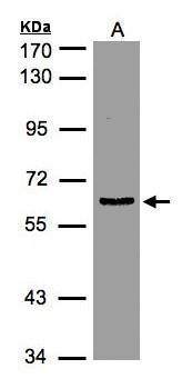
Sample(30 μg of whole cell lysate) A:HeLa S3 7.5% SDS PAGE diluted at 1:1000Application
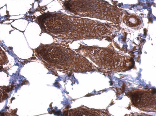
EIF3D antibody detects EIF3D protein at cytosol on mouse skin by immunohistochemical analysis. Sample: Paraffin-embedded mouse skin. EIF3D antibody dilution: 1:500.Application
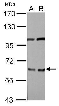
EIF3D antibody detects EIF3D protein by Western blot analysis. A. 30 µg PC-12 whole cell lysate/extract B. 30 µg Rat2 whole cell lysate/extract 7.5 % SDS-PAGE EIF3D antibody dilution: 1:3000Application
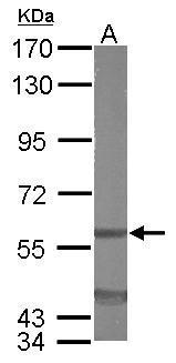
Sample (50 ug of whole cell lysate) A: mouse liver 7.5% SDS PAGE diluted at 1:5000Application
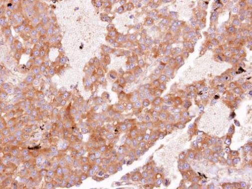
Immunohistochemical analysis of paraffin-embedded HUH7 xenograft, using EIF3 zeta antibody at 1:100 dilution.| Product Name | EIF3D antibody |
|---|---|
| Antibody Type | Primary Antibodies |
| Product description | Rabbit Polyclonal antibody to EIF3D (eukaryotic translation initiation factor 3, subunit D)Eukaryotic translation initiation factor-3 (eIF3), the largest of the eIFs, is a multiprotein complex composed of at least ten nonidentical subunits. The complex binds to the 40S ribosome and helps maintain the 40S and 60S ribosomal subunits in a dissociated state. It is also thought to play a role in the formation of the 40S initiation complex by interacting with the ternary complex of eIF2/GTP/methionyl-tRNA, and by promoting mRNA binding. The protein encoded by this gene is the major RNA binding subunit of the eIF3 complex. [provided by RefSeq] |
| Immunogen | Recombinant protein encompassing a sequence within the center region of human EIF3D. The exact sequence is proprietary. |
| Clonality | Polyclonal |
|---|---|
| Isotype | IgG |
| Host Species | Rabbit |
| Tested Applications | IHC-PWB |
| *Optimal dilutions/concentrations should be determined by the researcher. | |
| Species Reactivity | HumanMouseRat |
| Predicted Reactivity | Zebrafish, Xenopus laevis, Pig, Rhesus Monkey, Chimpanzee, BovineZebrafish (92%), Xenopus laevis (91%), Pig (99%), Rhesus Monkey (100%), Chimpanzee (100%), Bovine (100%) |
| Concentration | 1mg/ml |
| Purification | Affinity purified |
| Gene Synonyms | EIF3S7 antibody, MGC126526 antibody, MGC17258 antibody, eIF3-p66 antibody, eIF3-zeta antibody, EIF3D antibody, translation initiation factor eIF3 p66 subunit antibody, eukaryotic translation initiation factor 3 subunit D antibody, eukaryotic translation in |
|---|---|
| Molecular Weight(MW) | 64 kDa (note) |
| Cellular Localization | Cytoplasm |
Application
Sample(30 μg of whole cell lysate) A:HeLa S3 7.5% SDS PAGE diluted at 1:1000
Application
EIF3D antibody detects EIF3D protein at cytosol on mouse skin by immunohistochemical analysis. Sample: Paraffin-embedded mouse skin. EIF3D antibody dilution: 1:500.
Application
EIF3D antibody detects EIF3D protein by Western blot analysis. A. 30 µg PC-12 whole cell lysate/extract B. 30 µg Rat2 whole cell lysate/extract 7.5 % SDS-PAGE EIF3D antibody dilution: 1:3000
Application
Sample (50 ug of whole cell lysate) A: mouse liver 7.5% SDS PAGE diluted at 1:5000
Application
Immunohistochemical analysis of paraffin-embedded HUH7 xenograft, using EIF3 zeta antibody at 1:100 dilution.| Application Notes | *Optimal dilutions/concentrations should be determined by the researcher. |
|---|
| Form | Liquid |
|---|---|
| Storage Instructions | Keep as concentrated solution. Aliquot and store at -20ºC or below. Avoid multiple freeze-thaw cycles. |
| Storage Buffer | 0.1M Tris, 0.1M Glycine, 10% Glycerol (pH7). 0.01% Thimerosal was added as a preservative. |


