Application
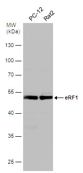
Various whole cell extracts (30 μg) were separated by 10% SDS-PAGE, and the membrane was blotted with eRF1 antibody diluted at 1:500.Application
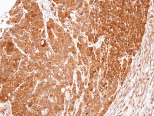
Immunohistochemical analysis of paraffin-embedded SW480 xenograft, using eRF1 antibody at 1:100 dilution.Application
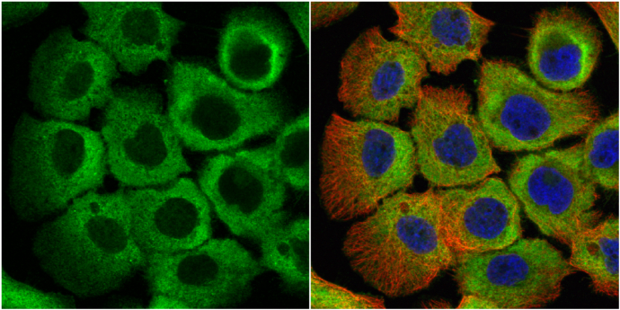
eRF1 antibody detects eRF1 protein at cytoplasm by immunofluorescent analysis. Sample: A431 cells were fixed in 4% paraformaldehyde at RT for 15 min. Green: eRF1 protein stained by eRF1 antibody diluted at 1:500. Red: alpha Tubulin, a cytoskeleton marker, stained by alpha Tubulin antibody [GT114] diluted at 1:1000. Blue: Hoechst 33342 staining.Application
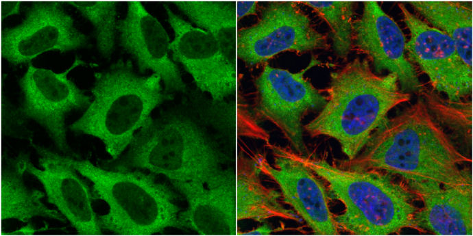
eRF1 antibody detects eRF1 protein at cytoplasm by immunofluorescent analysis. Sample: HeLa cells were fixed in 4% paraformaldehyde at RT for 15 min. Green: eRF1 protein stained by eRF1 antibody diluted at 1:500. Red: phalloidin, a cytoskeleton marker, stained by phalloidin (invitrogen, A12380) diluted at 1:200. Blue: Hoechst 33342 staining.Application
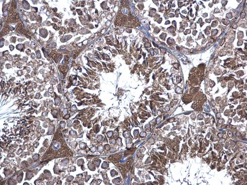
eRF1 antibody detects eRF1 protein at cytoplasm on mouse testis by immunohistochemical analysis. Sample: Paraffin-embedded mouse testis. eRF1 antibody diluted at 1:500.Application
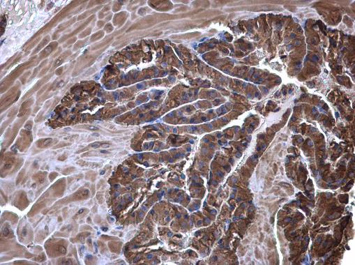
eRF1 antibody detects eRF1 protein at cytoplasm on mouse prostate by immunohistochemical analysis. Sample: Paraffin-embedded mouse prostate. eRF1 antibody diluted at 1:500.| Product Name | eRF1 antibody |
|---|---|
| Antibody Type | Primary Antibodies |
| Product description | Rabbit Polyclonal antibody to eRF1 (eukaryotic translation termination factor 1)Termination of protein biosynthesis and release of the nascent polypeptide chain are signaled by the presence of an in-frame stop codon at the aminoacyl site of the ribosome. The process of translation termination is universal and is mediated by protein release factors (RFs) and GTP. A class 1 RF recognizes the stop codon and promotes the hydrolysis of the ester bond linking the polypeptide chain with the peptidyl site tRNA, a reaction catalyzed at the peptidyl transferase center of the ribosome. Class 2 RFs, which are not codon specific and do not recognize codons, stimulate class 1 RF activity and confer GTP dependency upon the process. In prokaryotes, both class 1 RFs, RF1 and RF2, recognize UAA; however, UAG and UGA are decoded specifically by RF1 and RF2, respectively. In eukaryotes, eRF1, or ETF1, the functional counterpart of RF1 and RF2, functions as an omnipotent RF, decoding all 3 stop codons (Frolova et al., 1994 [PubMed 7990965]).[supplied by OMIM] |
| Immunogen | Carrier-protein conjugated synthetic peptide encompassing a sequence within the C-terminus region of human eRF1. The exact sequence is proprietary. |
| Clonality | Polyclonal |
|---|---|
| Isotype | IgG |
| Host Species | Rabbit |
| Tested Applications | ICC/IFIHC-PWB |
| *Optimal dilutions/concentrations should be determined by the researcher.
: |
|
| Species Reactivity | HumanMouseRat |
| Predicted Reactivity | Dog, Rabbit, Rhesus Monkey, BovineDog (100%), Rabbit (100%), Rhesus Monkey (100%), Bovine (100%) |
| Concentration | 1mg/ml |
| Purification | Affinity purified |
| Gene Synonyms | D5S1995 antibody ERF antibody ERF1 antibody MGC111066 antibody RF1 antibody SUP45L1 antibody TB3-1 antibody ETF1 antibody eukaryotic release factor 1 antibody eukaryotic peptide chain release factor subunit 1 antibody polypeptide chain release fa |
|---|---|
| Molecular Weight(MW) | 49 kDa (note) (5) |
| Cellular Localization | Cytoplasm |
Application
Various whole cell extracts (30 μg) were separated by 10% SDS-PAGE, and the membrane was blotted with eRF1 antibody diluted at 1:500.
Application
Immunohistochemical analysis of paraffin-embedded SW480 xenograft, using eRF1 antibody at 1:100 dilution.
Application
eRF1 antibody detects eRF1 protein at cytoplasm by immunofluorescent analysis. Sample: A431 cells were fixed in 4% paraformaldehyde at RT for 15 min. Green: eRF1 protein stained by eRF1 antibody diluted at 1:500. Red: alpha Tubulin, a cytoskeleton marker, stained by alpha Tubulin antibody [GT114] diluted at 1:1000. Blue: Hoechst 33342 staining.
Application
eRF1 antibody detects eRF1 protein at cytoplasm by immunofluorescent analysis. Sample: HeLa cells were fixed in 4% paraformaldehyde at RT for 15 min. Green: eRF1 protein stained by eRF1 antibody diluted at 1:500. Red: phalloidin, a cytoskeleton marker, stained by phalloidin (invitrogen, A12380) diluted at 1:200. Blue: Hoechst 33342 staining.
Application
eRF1 antibody detects eRF1 protein at cytoplasm on mouse testis by immunohistochemical analysis. Sample: Paraffin-embedded mouse testis. eRF1 antibody diluted at 1:500.
Application
eRF1 antibody detects eRF1 protein at cytoplasm on mouse prostate by immunohistochemical analysis. Sample: Paraffin-embedded mouse prostate. eRF1 antibody diluted at 1:500.| Positive Control | Neuro 2A , C8D30 , NIH-3T3 , Raw264.7 , C2C12 , PC-12 , Rat2 |
|---|---|
| Application Notes | *Optimal dilutions/concentrations should be determined by the researcher.
: |
| Form | Liquid |
|---|---|
| Storage Instructions | Keep as concentrated solution. Aliquot and store at -20ºC or below. Avoid multiple freeze-thaw cycles. |
| Storage Buffer | 0.1M Tris, 0.1M Glycine, 10% Glycerol (pH7). 0.01% Thimerosal was added as a preservative. |


