Application
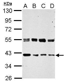
Sample (30 ug of whole cell lysate) A: 293T B: A431 C: HeLa D: HepG2 10% SDS PAGE diluted at 1:1000Application
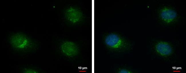
ERGIC3 antibody detects ERGIC3 protein at cytoplasm by immunofluorescent analysis. Sample: HeLa cells were fixed in 4% paraformaldehyde at RT for 15 min. Green: ERGIC3 protein stained by ERGIC3 antibody diluted at 1:1000. Blue: Hoechst 33342 staining.Application
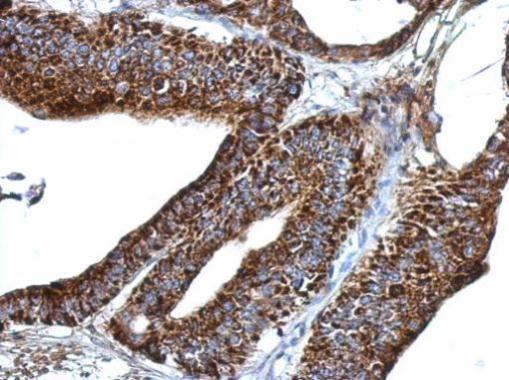
Immunohistochemical analysis of paraffin-embedded human gastric cancer, using ERGIC3 antibody at 1:500 dilution.| Product Name | ERGIC3 antibody |
|---|---|
| Antibody Type | Primary Antibodies |
| Product description | Rabbit Polyclonal antibody to ERGIC3 (endoplasmic reticulum-Golgi intermediate compartment protein 3) |
| Immunogen | Recombinant protein encompassing a sequence within the center region of human ERGIC3. The exact sequence is proprietary. |
| Clonality | Polyclonal |
|---|---|
| Isotype | IgG |
| Host Species | Rabbit |
| Tested Applications | ICC/IFIHC-PWB |
| *Optimal dilutions/concentrations should be determined by the researcher.
: |
|
| Species Reactivity | Human |
| Predicted Reactivity | Mouse, Rat, Zebrafish, Xenopus laevis, Rabbit, BovineMouse (96%), Rat (97%), Zebrafish (86%), Xenopus laevis (83%), Rabbit (97%), Bovine (96%) |
| Concentration | 1mg/ml |
| Purification | Affinity purified |
| Gene Synonyms | C20orf47 antibody CGI-54 antibody Erv46 antibody NY-BR-84 antibody PRO0989 antibody SDBCAG84 antibody dJ477O4.2 antibody ERGIC3 antibody serologically defined breast cancer antigen NY-BR-84 antibody serologically defined breast cancer antigen 84 a |
|---|---|
| Molecular Weight(MW) | 43 kDa (note) (3) |
| Cellular Localization | Endoplasmic reticulum-Golgi intermediate compartment membrane; Multi-pass membrane protein , Golgi apparatus , cis-Golgi network membrane; Multi-pass membrane protein , Endoplasmic reticulum membrane; Multi-pass membrane protein |
Application
Sample (30 ug of whole cell lysate) A: 293T B: A431 C: HeLa D: HepG2 10% SDS PAGE diluted at 1:1000
Application
ERGIC3 antibody detects ERGIC3 protein at cytoplasm by immunofluorescent analysis. Sample: HeLa cells were fixed in 4% paraformaldehyde at RT for 15 min. Green: ERGIC3 protein stained by ERGIC3 antibody diluted at 1:1000. Blue: Hoechst 33342 staining.
Application
Immunohistochemical analysis of paraffin-embedded human gastric cancer, using ERGIC3 antibody at 1:500 dilution.| Positive Control | 293T , A431 , HeLa , HepG2 |
|---|---|
| Application Notes | *Optimal dilutions/concentrations should be determined by the researcher.
: |
| Form | Liquid |
|---|---|
| Storage Instructions | Keep as concentrated solution. Aliquot and store at -20ºC or below. Avoid multiple freeze-thaw cycles. |
| Storage Buffer | 1XPBS, 20% Glycerol (pH7). 0.01% Thimerosal was added as a preservative. |


