Application
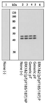
Extracts prepared from PC12 cells left unstimulated (1) or stimulated with sorbitol (2-6) were resolved by SDS-PAGE on a 10% polyacrylamide gel and transferred to PVDF. Membranes were blocked with a 5% BSA-TBST buffer overnight at 4 degree C then incubated withApplication
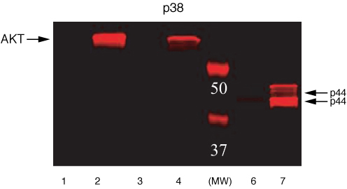
NIH3T3 or NIH3T3-L1 cells were treated with either PDGF or Insulin. Western Blots were performed using either Akt [pS473] or Erk [pTpY185/187] primary antibodies. Anti-rabbit secondary antibody conjugated to Alexa fluor 680 was used for detection. Akt [pS473]: Lane 1 – NIH3T3 control, 20 µg Lane 2 – NIH3T3 + PDGF, 20 µg Lane 3 – NIH3T3-L1 control, 20 µg Lane 4 – NIH3T3-L1 + Insulin, 20 µg. ERK [pTpY185/187]: Lane 6 – NIH3T3 control, 5 µg Lane 7 – NIH3T3 + PDGF, 5 µg| Product Name | ERK1/2 (phospho Thr185/Tyr187) antibody |
|---|---|
| Antibody Type | Primary Antibodies |
| Product description | Rabbit polyclonal antibody to ERK1/2 (phospho Thr185/Tyr187)ERK (extracellular signal-regulated kinase), also known as MAPK (mitogen-activated protein kinase) has two closely related isoforms of 44 kDa and 42 kDa, respectively. These kinases belong to a family of serine/threonine kinases that are activated upon treatment of cells with a large variety of stimuli including mitogens, hormones, growth factors, cytokines, and bioactive peptides. Cell stimulation induces the activation of a signalling cascade, the downstream effects of which have been linked to the regulation of cell growth and differentiation as well as the cytoskeleton. ERK 1 and ERK 2 are fully activated by phosphorylation within a Thr-Glu-Tyr motif of the activation loop on both a threonine and a tyrosine residue by MEKs (MAPK/ERK kinases), thereby greatly elevating the activity of the ERK 1 + 2 enzymes. |
| Immunogen | Synthetic peptide (Human) derived from the region of ERK 1 + 2 that contains threonine 202/185 and tyrosine 204/187, based on the human sequence. This region is conserved among many species including rat, mouse, cow, frog, snail, nematode, and fruit fly. |
| Modification | p-Thr185/Tyr187 |
| Clonality | Polyclonal |
|---|---|
| Isotype | IgG |
| Host Species | Rabbit |
| Tested Applications | WB |
| WB: Use at a dilution of 1/1000. Predicted molecular weight: 44,42 kDa. Can be blocked with GTX25354 NIH3T3 whole cell lysate, + / - PDGF stimulation (pair) , GTX25313 PC12 whole cell lysate, + / - Sorbitol treatment or GTX25255 ERK 1 + 2 phospho T185 / 202 + Y187 / 204 and non-phospho peptides (pair) Optimal dilutions/concentrations should be determined by the end user.: |
|
| Species Reactivity | HumanMouse |
| Concentration | 1mg/ml |
| Purification | Affinity purified |
| Gene Synonyms | ERK1 & ERK2 176982 PKC2 116590 Erk2 5590 Q05513 Mapk1 PRKCZ ERK1 (phospho T185/T202) ERK1 (phospho Thr185/Thr202) ERK1 (pT185/pT202) ERK1 (pThr185/pThr202) ERK1 phospho T185/T202 ERK1 phospho Thr185/Thr202 Phospho ERK1 (T185/T202) Phospho E |
|---|---|
| Target | ERK1/2 (phospho Thr185/Tyr187) |
| Tissue Specificity | The sequence of the immunogenic peptide used to generate includes the amino acids found in the region of both ERK1 and ERK2 including specific threonine and tyrosine residues. In ERK1, these residues are at positions 185 and 187, respectively. In ERK2, these same residues are actually at positions 202 and 204, respectively of the native protein. The antibody recognizes the same sequence in both proteins (ERK1/2) as long as the T and the Y are phosphorylated. Does not recognize either monophospho- forms of ERK1/2 or the non-phosphorylated proteins. |
Application
Extracts prepared from PC12 cells left unstimulated (1) or stimulated with sorbitol (2-6) were resolved by SDS-PAGE on a 10% polyacrylamide gel and transferred to PVDF. Membranes were blocked with a 5% BSA-TBST buffer overnight at 4 degree C then incubated with
Application
NIH3T3 or NIH3T3-L1 cells were treated with either PDGF or Insulin. Western Blots were performed using either Akt [pS473] or Erk [pTpY185/187] primary antibodies. Anti-rabbit secondary antibody conjugated to Alexa fluor 680 was used for detection. Akt [pS473]: Lane 1 – NIH3T3 control, 20 µg Lane 2 – NIH3T3 + PDGF, 20 µg Lane 3 – NIH3T3-L1 control, 20 µg Lane 4 – NIH3T3-L1 + Insulin, 20 µg. ERK [pTpY185/187]: Lane 6 – NIH3T3 control, 5 µg Lane 7 – NIH3T3 + PDGF, 5 µg| Positive Control | PC12 cells +/- Sorbitol |
|---|---|
| Application Notes | WB: Use at a dilution of 1/1000. Predicted molecular weight: 44,42 kDa. Can be blocked with GTX25354 NIH3T3 whole cell lysate, + / - PDGF stimulation (pair) , GTX25313 PC12 whole cell lysate, + / - Sorbitol treatment or GTX25255 ERK 1 + 2 phospho T185 / 202 + Y187 / 204 and non-phospho peptides (pair) Optimal dilutions/concentrations should be determined by the end user.: |
| Form | Liquid |
|---|---|
| Storage Instructions | Keep as concentrated solution. Store at 4ºC short term. For extended storage aliquot and store at -20ºC or below. Avoid freeze-thaw cycles. |
| Storage Buffer | Preservative: 0.05% Sodium Azide; Constituents: 50% Glycerol, PBS, 1mg/ml BSA (IgG, protease free). pH 7.3 |


