Application
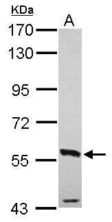
Sample (30 ug of whole cell lysate) A: NT2D1 7.5% SDS PAGE diluted at 1:1000Application
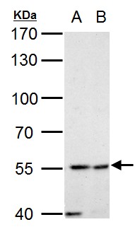
Estrogen Receptor beta antibody detects Estrogen Receptor beta protein by western blot analysis. A. 30 μg MCF-7 whole cell lysate/extract B. 30 μg MDA-MB-231 whole cell lysate/extract 7.5 % SDS-PAGE Estrogen Receptor beta antibody dilution: 1:1000Application
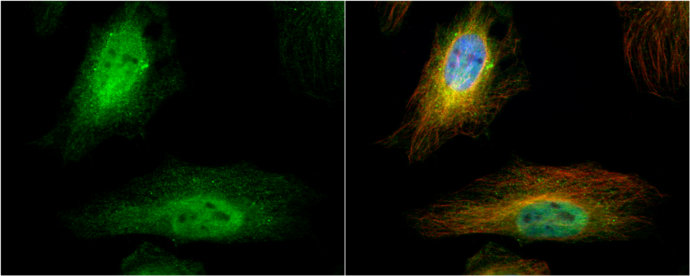
Estrogen Receptor beta antibody detects Estrogen Receptor beta protein at cytoplasm and nucleus by immunofluorescent analysis. Sample: HeLa cells were fixed in 4% paraformaldehyde at RT for 15 min. Green: Estrogen Receptor beta protein stained by Estrogen Receptor beta antibody diluted at 1:500. Red: alpha Tubulin, a cytoskeleton marker, stained by alpha Tubulin antibody [B-5-1-2] diluted at 1:10000. Blue: Hoechst 33342 staining.Application
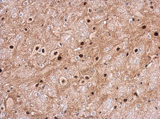
Estrogen Receptor beta antibody detects Estrogen Receptor beta protein at cytoplasm and nucleus in mouse brain by immunohistochemical analysis. Sample: Paraffin-embedded mouse brain. Estrogen Receptor beta antibody diluted at 1:200.WB
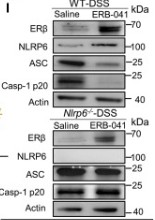
(I) ERβ, NLRP6, ASC, and Casp-1 p20 expression of mice colon homogenate. See also Figure S1 and Data S1.WB
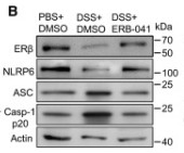
(B) Immunoblotting of treated cell lysates. Data are representative of three independent experiments. ∗p < 0.05, ∗∗p < 0.01, by two-way ANOVA with Tukey’s post hoc analysis. Normal NCM-460 cells were stimulated with the selective ERβ agonist (ERB-041, 1 μM) and ERα agonist (PPT, 1 μM) for 48 h.IHC
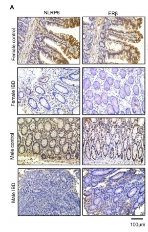
(A) Immunohistochemistry analysis of NLRP6 and ERβ in different intestinal tissue sections. (Scale bars, 100 µm).WB
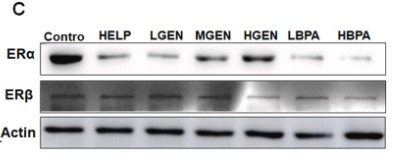
(C) Protein expression levels of ERα and ERβ in the liver tissue.WB
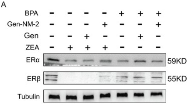
(A) Expression levels of ERα and ERβ.WB
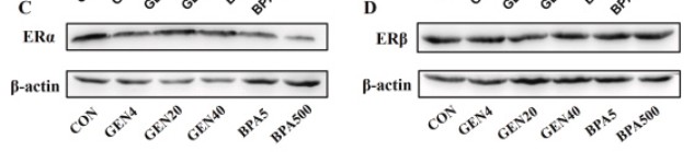
(C and D) Western blot analysis of ERα and ERβ.WB
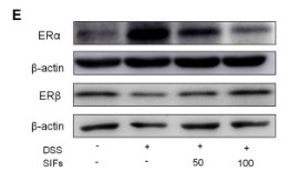
(E) Protein levels of ERα in the colon tissues. The relative protein expression of ERα was normalized to β-actin.IHC
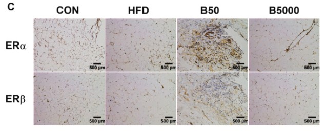
(C) ERα and ERβ in AFT were located by IHC. Bars: 500 µm.| Product Name | Estrogen Receptor beta antibody |
|---|---|
| Antibody Type | Primary Antibodies |
| Product description | Rabbit Polyclonal antibody to Estrogen Receptor beta (estrogen receptor 2 (ER beta))This gene encodes a member of the family of estrogen receptors and superfamily of nuclear receptor transcription factors. The gene product contains an N-terminal DNA binding domain and C-terminal ligand binding domain and is localized to the nucleus, cytoplasm, and mitochondria. Upon binding to 17beta-estradiol or related ligands, the encoded protein forms homo- or hetero-dimers that interact with specific DNA sequences to activate transcription. Some isoforms dominantly inhibit the activity of other estrogen receptor family members. Several alternatively spliced transcript variants of this gene have been described, but the full-length nature of some of these variants has not been fully characterized. [provided by RefSeq] |
| Immunogen | Recombinant protein encompassing a sequence within the center region of human Estrogen Receptor beta. The exact sequence is proprietary. |
| Modification | Acetylated |
| Clonality | Polyclonal |
|---|---|
| Isotype | IgG |
| Host Species | Rabbit |
| Tested Applications | ICC/IFIHCIHC-PWB |
| WB:1:500~1:2000 ICC/IF:1:100~1:500 IHC-P:1:50~1:200 |
|
| Species Reactivity | HumanMouse |
| Predicted Reactivity | Pig, Sheep, Rhesus Monkey, BovinePig (82%), Sheep (81%), Rhesus Monkey (93%), Bovine (81%) |
| Concentration | 0.5mg/ml |
| Purification | Affinity purified |
| Gene Synonyms | ER-BETA antibody ESR-BETA antibody ESRB antibody ESTRB antibody Erb antibody NR3A2 antibody ESR2 antibody estrogen nuclear receptor beta variant a antibody estrogen nuclear receptor beta variant b antibody estrogen receptor beta antibody estrogen |
|---|---|
| Molecular Weight(MW) | 59 kDa (note) (9) |
| Cellular Localization | Nucleus |
Application
Sample (30 ug of whole cell lysate) A: NT2D1 7.5% SDS PAGE diluted at 1:1000
Application
Estrogen Receptor beta antibody detects Estrogen Receptor beta protein by western blot analysis. A. 30 μg MCF-7 whole cell lysate/extract B. 30 μg MDA-MB-231 whole cell lysate/extract 7.5 % SDS-PAGE Estrogen Receptor beta antibody dilution: 1:1000
Application
Estrogen Receptor beta antibody detects Estrogen Receptor beta protein at cytoplasm and nucleus by immunofluorescent analysis. Sample: HeLa cells were fixed in 4% paraformaldehyde at RT for 15 min. Green: Estrogen Receptor beta protein stained by Estrogen Receptor beta antibody diluted at 1:500. Red: alpha Tubulin, a cytoskeleton marker, stained by alpha Tubulin antibody [B-5-1-2] diluted at 1:10000. Blue: Hoechst 33342 staining.
Application
Estrogen Receptor beta antibody detects Estrogen Receptor beta protein at cytoplasm and nucleus in mouse brain by immunohistochemical analysis. Sample: Paraffin-embedded mouse brain. Estrogen Receptor beta antibody diluted at 1:200.
WB
(I) ERβ, NLRP6, ASC, and Casp-1 p20 expression of mice colon homogenate. See also Figure S1 and Data S1.
WB
(B) Immunoblotting of treated cell lysates. Data are representative of three independent experiments. ∗p < 0.05, ∗∗p < 0.01, by two-way ANOVA with Tukey’s post hoc analysis. Normal NCM-460 cells were stimulated with the selective ERβ agonist (ERB-041, 1 μM) and ERα agonist (PPT, 1 μM) for 48 h.
IHC
(A) Immunohistochemistry analysis of NLRP6 and ERβ in different intestinal tissue sections. (Scale bars, 100 µm).
WB
(C) Protein expression levels of ERα and ERβ in the liver tissue.
WB
(A) Expression levels of ERα and ERβ.
WB
(C and D) Western blot analysis of ERα and ERβ.
WB
(E) Protein levels of ERα in the colon tissues. The relative protein expression of ERα was normalized to β-actin.
IHC
(C) ERα and ERβ in AFT were located by IHC. Bars: 500 µm.| Positive Control | NT2D1 , IMR32 , U87-MG , MCF-7 , MDA-MB-231 |
|---|---|
| Application Notes | WB:1:500~1:2000 ICC/IF:1:100~1:500 IHC-P:1:50~1:200 |
| Form | Liquid |
|---|---|
| Storage Instructions | Keep as concentrated solution. Aliquot and store at -20ºC or below. Avoid multiple freeze-thaw cycles. |
| Storage Buffer | 0.1M Tris, 0.1M Glycine, 20% Glycerol (pH7). 0.01% Thimerosal was added as a preservative. |


