Application
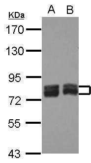
Sample (30 µg of whole cell lysate) A: HeLa B: NT2D1 7.5% SDS PAGE diluted at 1:1000 The HRP-conjugated anti-rabbit IgG antibody was used to detect the primary antibody.Application
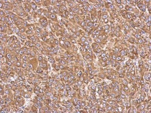
Immunohistochemical analysis of paraffin-embedded HBL435 xenograft, using FACL4 antibody at 1:500 dilution.Application
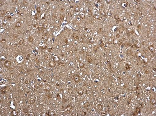
FACL4 antibody [C3], C-term detects FACL4 protein at cytosol on rat middle brain by immunohistochemical analysis. Sample: Paraffin-embedded rat middle brain. FACL4 antibody [C3], C-term dilution: 1:500.Application
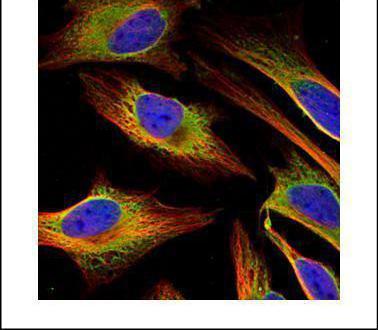
Confocal immunofluorescence analysis (Olympus FV10i) of methanol-fixed HeLa, using FACL4 antibody (Green) at 1:500 dilution. Alpha-tubulin filaments were labeled with (Red) at 1:2000.Application
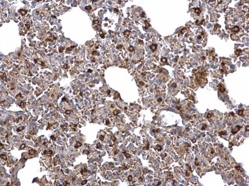
FACL4 antibody [C3], C-term detects FACL4 protein at cytosol on mouse lymph node by immunohistochemical analysis. Sample: Paraffin-embedded mouse lymph node. FACL4 antibody [C3], C-term dilution: 1:500.Application
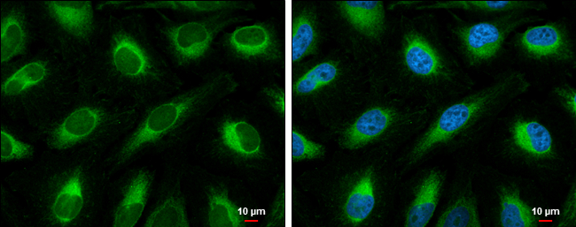
FACL4 antibody [C3], C-term detects FACL4 protein at endoplasmic reticulum by immunofluorescent analysis. Sample: HeLa cells were fixed in 4% paraformaldehyde at RT for 15 min. Green: FACL4 protein stained by FACL4 antibody [C3], C-term diluted at 1:500. Blue: Hoechst 33342 staining. Scale bar = 10 μm.Application
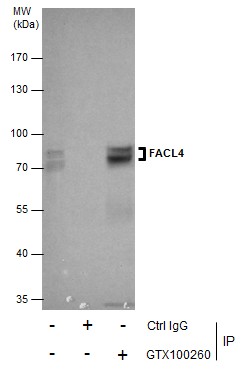
Immunoprecipitation of FACL4 protein from HeLa whole cell extracts using 5 μg of FACL4 antibody [C3], C-term . Western blot analysis was performed using FACL4 antibody [C3], C-term . EasyBlot anti-Rabbit IgG was used as a secondary reagent.Application
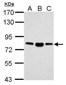
Sample (30 µg of whole cell lysate) A: NIH-3T3 B: JC C: BCL-1 7.5% SDS PAGE diluted at 1:5000 The HRP-conjugated anti-rabbit IgG antibody was used to detect the primary antibody.| Product Name | FACL4 antibody , C-term |
|---|---|
| Antibody Type | Primary Antibodies |
| Product description | Rabbit Polyclonal antibody to FACL4 (acyl-CoA synthetase long-chain family member 4)The protein encoded by this gene is an isozyme of the long-chain fatty-acid-coenzyme A ligase family. Although differing in substrate specificity, subcellular localization, and tissue distribution, all isozymes of this family convert free long-chain fatty acids into fatty acyl-CoA esters, and thereby play a key role in lipid biosynthesis and fatty acid degradation. This isozyme preferentially utilizes arachidonate as substrate. The absence of this enzyme may contribute to the mental retardation or Alport syndrome. Alternative splicing of this gene generates 2 transcript variants. [provided by RefSeq] |
| Immunogen | Carrier-protein conjugated synthetic peptide encompassing a sequence within the C-terminus region of human FACL4. The exact sequence is proprietary. |
| Clonality | Polyclonal |
|---|---|
| Isotype | IgG |
| Host Species | Rabbit |
| Tested Applications | ICC/IFIHC-PIPWB |
| *Optimal dilutions/concentrations should be determined by the researcher.
: |
|
| Species Reactivity | HumanMouseRat |
| Predicted Reactivity | Pig, Rhesus MonkeyPig (100%), Rhesus Monkey (100%) |
| Concentration | 1mg/ml (Please refer to the vial label for the specific concentration) |
| Purification | Affinity purified |
| Gene Synonyms | ACS4 antibody FACL4 antibody LACS4 antibody MRX63 antibody MRX68 antibody ACSL4 antibody acyl-CoA synthetase 4 antibody long-chain acyl-CoA synthetase 4 antibody lignoceroyl-CoA synthase antibody LACS 4 antibody long-chain fatty-acid-Coenzyme A l |
|---|---|
| Molecular Weight(MW) | 79 kDa (note) (4) |
| Cellular Localization | Mitochondrion outer membrane; Single-pass type III membrane protein , Peroxisome membrane; Single-pass type III membrane protein , Microsome membrane; Single-pass type III membrane protein , Endoplasmic reticulum membrane; Single-pass type III membrane pr |
Application
Sample (30 µg of whole cell lysate) A: HeLa B: NT2D1 7.5% SDS PAGE diluted at 1:1000 The HRP-conjugated anti-rabbit IgG antibody was used to detect the primary antibody.
Application
Immunohistochemical analysis of paraffin-embedded HBL435 xenograft, using FACL4 antibody at 1:500 dilution.
Application
FACL4 antibody [C3], C-term detects FACL4 protein at cytosol on rat middle brain by immunohistochemical analysis. Sample: Paraffin-embedded rat middle brain. FACL4 antibody [C3], C-term dilution: 1:500.
Application
Confocal immunofluorescence analysis (Olympus FV10i) of methanol-fixed HeLa, using FACL4 antibody (Green) at 1:500 dilution. Alpha-tubulin filaments were labeled with (Red) at 1:2000.
Application
FACL4 antibody [C3], C-term detects FACL4 protein at cytosol on mouse lymph node by immunohistochemical analysis. Sample: Paraffin-embedded mouse lymph node. FACL4 antibody [C3], C-term dilution: 1:500.
Application
FACL4 antibody [C3], C-term detects FACL4 protein at endoplasmic reticulum by immunofluorescent analysis. Sample: HeLa cells were fixed in 4% paraformaldehyde at RT for 15 min. Green: FACL4 protein stained by FACL4 antibody [C3], C-term diluted at 1:500. Blue: Hoechst 33342 staining. Scale bar = 10 μm.
Application
Immunoprecipitation of FACL4 protein from HeLa whole cell extracts using 5 μg of FACL4 antibody [C3], C-term . Western blot analysis was performed using FACL4 antibody [C3], C-term . EasyBlot anti-Rabbit IgG was used as a secondary reagent.
Application
Sample (30 µg of whole cell lysate) A: NIH-3T3 B: JC C: BCL-1 7.5% SDS PAGE diluted at 1:5000 The HRP-conjugated anti-rabbit IgG antibody was used to detect the primary antibody.| Positive Control | HeLa , NT2D1 , NIH-3T3 , JC , BCL-1 |
|---|---|
| Application Notes | *Optimal dilutions/concentrations should be determined by the researcher.
: |
| Form | Liquid |
|---|---|
| Storage Instructions | Keep as concentrated solution. Aliquot and store at -20ºC or below. Avoid multiple freeze-thaw cycles. |
| Storage Buffer | 1XPBS, 1% BSA, 20% Glycerol (pH7). 0.01% Thimerosal was added as a preservative. |


