Application
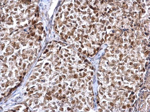
FEN1 antibody detects FEN1 protein at nucleus on human breast carcinoma by immunohistochemical analysis. Sample: Paraffin-embedded human breast carcinoma. FEN1 antibody diluted at 1:500.Application
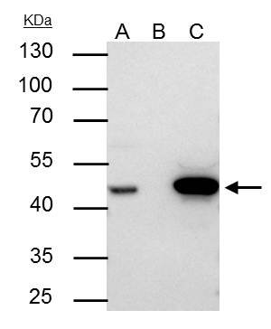
FEN1 antibody immunoprecipitates FEN1 protein in IP experiments. IP samples: Jurkat whole cell extract A. 30 μg Jurkat whole cell extract B. Control with 4 μg of preimmune Rabbit IgG C. Immunoprecipitation of FEN1 protein by 4 μg FEN1 antibody 10 % SDS-PAGE The immunoprecipitated FEN1 protein was detected by FEN1 antibody diluted at 1:500. [EasyBlot anti-rabbit IgG was used as a secondary reagent]Application
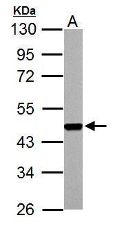
FEN1 antibody detects FEN1 protein by western blot analysis. A. 30 µg PC-12 whole cell lysate/extract 10% SDS-PAGE FEN1 antibody dilution: 1:1000 The HRP-conjugated anti-rabbit IgG antibody was used to detect the primary antibody.Application
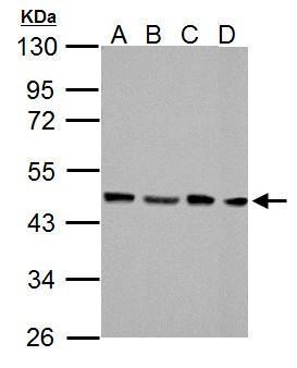
FEN1 antibody detects FEN1 protein by western blot analysis. A. 30 µg NIH-3T3 whole cell lysate/extract B. 30 µg JC whole cell lysate/extract C. 30 µg BCL-1 whole cell lysate/extract D. 30 µg Raw264.7 whole cell lysate/extract 10% SDS-PAGE FEN1 antibody dilution: 1:1000 The HRP-conjugated anti-rabbit IgG antibody was used to detect the primary antibody.Application
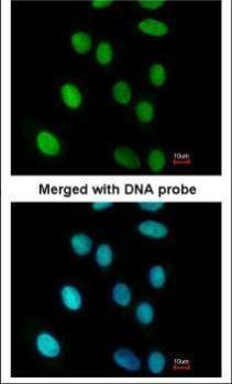
Immunofluorescence analysis of paraformaldehyde-fixed A549, using FEN1 antibody at 1:200 dilution.Application
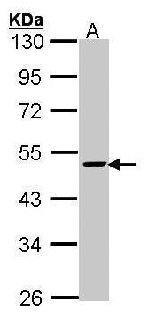
Sample (30 µg of whole cell lysate) A: HepG2 10% SDS PAGE diluted at 1:1000 The HRP-conjugated anti-rabbit IgG antibody was used to detect the primary antibody.| Product Name | FEN1 antibody |
|---|---|
| Antibody Type | Primary Antibodies |
| Product description | Rabbit Polyclonal antibody to FEN1 (flap structure-specific endonuclease 1)The protein encoded by this gene removes 5' overhanging flaps in DNA repair and processes the 5' ends of Okazaki fragments in lagging strand DNA synthesis. Direct physical interaction between this protein and AP endonuclease 1 during long-patch base excision repair provides coordinated loading of the proteins onto the substrate, thus passing the substrate from one enzyme to another. The protein is a member of the XPG/RAD2 endonuclease family and is one of ten proteins essential for cell-free DNA replication. DNA secondary structure can inhibit flap processing at certain trinucleotide repeats in a length-dependent manner by concealing the 5' end of the flap that is necessary for both binding and cleavage by the protein encoded by this gene. Therefore, secondary structure can deter the protective function of this protein, leading to site-specific trinucleotide expansions. [provided by RefSeq] |
| Immunogen | Recombinant protein encompassing a sequence within the center region of human FEN1. The exact sequence is proprietary. |
| Clonality | Polyclonal |
|---|---|
| Isotype | IgG |
| Host Species | Rabbit |
| Tested Applications | ICC/IFIHC-PIPWBPLA |
| *Optimal dilutions/concentrations should be determined by the researcher. | |
| Species Reactivity | HumanMouseRat |
| Predicted Reactivity | Sheep, Rhesus Monkey, BovineSheep (94%), Rhesus Monkey (98%), Bovine (96%) |
| Concentration | 1mg/ml |
| Purification | Affinity purified |
| Gene Synonyms | FEN-1 antibody, MF1 antibody, RAD2 antibody, FEN1 antibody, flap endonuclease 1 antibody, hFEN-1 antibody, maturation factor-1 antibody, DNase IV antibody, maturation factor 1 antibody, flap structure-specific endonuclease 1 antibody |
|---|---|
| Molecular Weight(MW) | 43 kDa (note) |
| Cellular Localization | Nucleus |
Application
FEN1 antibody detects FEN1 protein at nucleus on human breast carcinoma by immunohistochemical analysis. Sample: Paraffin-embedded human breast carcinoma. FEN1 antibody diluted at 1:500.
Application
FEN1 antibody immunoprecipitates FEN1 protein in IP experiments. IP samples: Jurkat whole cell extract A. 30 μg Jurkat whole cell extract B. Control with 4 μg of preimmune Rabbit IgG C. Immunoprecipitation of FEN1 protein by 4 μg FEN1 antibody 10 % SDS-PAGE The immunoprecipitated FEN1 protein was detected by FEN1 antibody diluted at 1:500. [EasyBlot anti-rabbit IgG was used as a secondary reagent]
Application
FEN1 antibody detects FEN1 protein by western blot analysis. A. 30 µg PC-12 whole cell lysate/extract 10% SDS-PAGE FEN1 antibody dilution: 1:1000 The HRP-conjugated anti-rabbit IgG antibody was used to detect the primary antibody.
Application
FEN1 antibody detects FEN1 protein by western blot analysis. A. 30 µg NIH-3T3 whole cell lysate/extract B. 30 µg JC whole cell lysate/extract C. 30 µg BCL-1 whole cell lysate/extract D. 30 µg Raw264.7 whole cell lysate/extract 10% SDS-PAGE FEN1 antibody dilution: 1:1000 The HRP-conjugated anti-rabbit IgG antibody was used to detect the primary antibody.
Application
Immunofluorescence analysis of paraformaldehyde-fixed A549, using FEN1 antibody at 1:200 dilution.
Application
Sample (30 µg of whole cell lysate) A: HepG2 10% SDS PAGE diluted at 1:1000 The HRP-conjugated anti-rabbit IgG antibody was used to detect the primary antibody.| Positive Control | HepG2 , NIH-3T3 , JC , BCL-1 , Raw264.7 , PC-12 |
|---|---|
| Application Notes | *Optimal dilutions/concentrations should be determined by the researcher. |
| Form | Liquid |
|---|---|
| Storage Instructions | Keep as concentrated solution. Aliquot and store at -20ºC or below. Avoid multiple freeze-thaw cycles. |
| Storage Buffer | 0.1M Tris, 0.1M Glycine, 10% Glycerol (pH7). 0.01% Thimerosal was added as a preservative. |


