Application
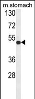
Western blot analysis of FERMT1 Antibody (Center) in mouse stomach tissue lysates (35ug/lane). FERMT1 (arrow) was detected using the purified PabApplication
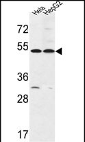
FERMT1 Antibody (Center) western blot analysis in Hela,HepG2 cell line lysates (35ug/lane).This demonstrates the FERMT1 antibody detected the FERMT1 protein (arrow).IF
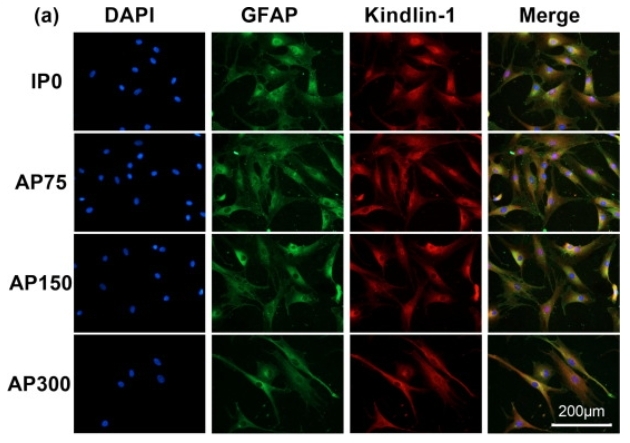
(a) Fluorescence staining of nuclei (blue color), GFAP (green color), and Kindlin-1 (red color) of astrocytes on different scaffolds.| Product Name | FERMT1 antibody, Internal |
|---|---|
| Antibody Type | Primary Antibodies |
| Product description | Rabbit polyclonal to FERMT1 (Internal)This gene encodes a member of the fermitin family, and contains a FERM domain and a pleckstrin homology domain. The encoded protein is involved in integrin signaling and linkage of the actin cytoskeleton to the extracellular matrix. Mutations in this gene have been linked to Kindler syndrome. [provided by RefSeq] |
| Immunogen | KLH conjugated synthetic peptide between 275~305 amino acids from the Center region of human FERMT1. |
| Clonality | Polyclonal |
|---|---|
| Isotype | IgG |
| Host Species | Rabbit |
| Tested Applications | ELISAFCIFIHCWB |
| ELISA: Use at 1:1,000 | |
| Species Reactivity | HumanMouseRat |
| Concentration | 0.2mg/ml |
| Purification | Affinity purified |
| Gene Synonyms | Unc112related protein 1, Kindlerin, Unc112related protein1, Kindlin-1, Kindlin1, FERMT1, Fermitin family homolog1, Kindlin syndrome protein, Unc-112-related protein 1, FERMT1, C20orf42, KIND1, URP1, Fermitin family homolog 1, Kindlin 1, Unc 112 related pro |
|---|---|
| Target | FERMT1 |
| Molecular Weight(MW) | 77.437 kDa (note) |
| Cellular Localization | Constituent of focal adhesions. Localized at the basal aspect of skin keratinocytes, close to the cell membrane. Colocalizes with filamentous actin. Upon TGFB1 treatment, it localizes to membrane ruffles. |
Application
Western blot analysis of FERMT1 Antibody (Center) in mouse stomach tissue lysates (35ug/lane). FERMT1 (arrow) was detected using the purified Pab
Application
FERMT1 Antibody (Center) western blot analysis in Hela,HepG2 cell line lysates (35ug/lane).This demonstrates the FERMT1 antibody detected the FERMT1 protein (arrow).
IF
(a) Fluorescence staining of nuclei (blue color), GFAP (green color), and Kindlin-1 (red color) of astrocytes on different scaffolds.| Application Notes | ELISA: Use at 1:1,000 |
|---|
| Form | Liquid |
|---|---|
| Storage Instructions | Store at at 2-8°C for up to 6 months. For long term storage, aliquot and store at -20 °C. Avoid repeated freeze/thaw cycles. |
| Storage Buffer | PBS with 0.09% (W/V) sodium azide |


