Application
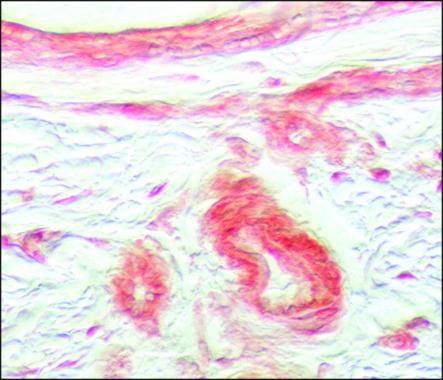
Staining of formalin-fixed, paraffin-embedded human umbilical cord sections with 5 μg/mL Anti-FGF Receptor 1 using Biotin-Anti-Rabbit IgG/ExtrAvidin®-PeroxidaseApplication
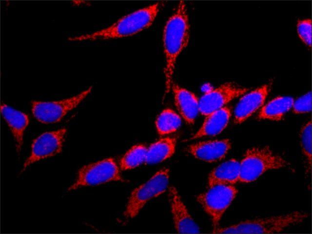
ICC/IF analysis of HeLa cells using FGFR1 antibody at a dilution of 1:100 (red) and DAPI (blue).| Product Name | FGFR1 antibody |
|---|---|
| Antibody Type | Primary Antibodies |
| Product description | Rabbit polyclonal to FGFR1Fibroblast growth factors (FGFs) are members of a large family of structurally related polypeptides (17-38 kDa) that are potent physiological regulators of growth and differentiation of a wide variety of cells of mesodermal, ectodermal and endodermal origin. FGFs are substantially involved in normal development, wound healing and repair, angiogenesis, a variety of neurotrophic activities, in hematopoiesis as well as in tissue remodeling and maintenance. They also have been implicated in pathological conditions such as tumorigenesis and metastasis. To date, the FGF family consists of at least 23 members designated FGF1 through FGF23. Four genes encoding for high affinity cell surface FGF receptors (FGFRs) have been identified: FGFR1 [flg-1(fms-like gene 1)]; FGFR2 [bek (bacterial expressed kinase gene product)]; FGFR3 (cek-2), and FGFR4. Multiple additional variants (isoforms) arising by alternative splicing have been reported: soluble, secreted, or possibly cleaved forms of FGFR1 and FGFR2 have also been found in body fluids or were artificially constructed, [e.g. a soluble FGF-binding protein containing the extracellular region of FGFR1 and the secreted form of placental alkaline phosphatase (FRAP1)]. FGFRs are members of the tyrosine kinase family of growth factor receptors. They are glycosylated 110- 150 kDa proteins that are constructed of an extracellular ligand binding region with either two (alpha type) or typically three (alpha type) immunoglbulin (Ig)-like domains and an eight amino acid acidic box, a transmembrane region, and a cytoplasmic split tyrosine kinase domain that is activated following ligand binding and receptor dimerization. The ligand binding site of FGFRs is confined to the extracellular Ig-like domains 2 and 3.FGFRs exhibit overlapping recognition and redundant specificity. One receptor type may bind with a similar affinity several of the FGFs. Also one FGF type may bind similarly to several distinct receptors. This accounts for the rather identical effects of different FGF ligands on common cell types. FGFis binding to cellular FGFRs depend on or is markedly facilitated by the low-affinity interaction of FGF with the polysaccharide component of the cell surface or extracellular matrix heparan sulfate proteoglycans (HSPG). For example, perlecan, a basement membrane HSPG, promotes high affinity binding of FGF2 in vitro and angiogenesis in vivo. Signal transduction by FGFRs requires dimerization or oligomerization and autophosphorylation of the receptors through their tyrosine kinase domain. Subsequent association with cytoplasmic signaling molecules leads to DNA synthesis or differentiation. The signaling and biological responses elicited by distinct FGFRs substantially differ and are dictated by the intracellular domain. At the mRNA level, FGFR1 is highly expressed in developing human tissues including the brain (preferentially in neurons), vascular basement membranes, skin, and bone growth plates. It may be found in most anchorage dependent cells on their membrane and also may be localized around and in nuclei. Pfeiffer syndrome, as well as other disorders of human skeletal development, is the result of a mutation in the extracellular domain of FGFR1. |
| Immunogen | Synthetic peptide: EALEERPAVMTSPLK conjugated to KLH, corresponding to amino acids 360-373 of Human FGFR1 with a C-terminally added lysine. |
| Clonality | Polyclonal |
|---|---|
| Isotype | IgG |
| Host Species | Rabbit |
| Tested Applications | ICC/IFIHC-PIPWB |
| For IHC-P (trypsin-digested): Use at a dilution of 1:200. For IP: Use at an assay dependent dilution. For WB: Use at a dilution of 1:400. The epitopes recognized by the antibody are resistant to routine formalin-fixation and paraffin embedding to other fixatives e.g. Methacam, Bouins solution, ethanol, and B5. Optimal dilutions/concentrations should be determined by the researcher.: |
|
| Species Reactivity | Human |
| Concentration | 1mg/ml |
| Purification | Affinity purified |
| Gene Synonyms | CEK CFGR N-SAM H5 NSAM P11362 FLT2 BFGFR 2260 FLG H3 H2 KAL2 H4 101600 FGFR1 C-FGR N SAM C FGR |
|---|---|
| Target | FGFR1 |
| Tissue Specificity | No reaction with FGFR-2 and FGFR-3 is detected. |
Application
Staining of formalin-fixed, paraffin-embedded human umbilical cord sections with 5 μg/mL Anti-FGF Receptor 1 using Biotin-Anti-Rabbit IgG/ExtrAvidin®-Peroxidase
Application
ICC/IF analysis of HeLa cells using FGFR1 antibody at a dilution of 1:100 (red) and DAPI (blue).| Application Notes | For IHC-P (trypsin-digested): Use at a dilution of 1:200. For IP: Use at an assay dependent dilution. For WB: Use at a dilution of 1:400. The epitopes recognized by the antibody are resistant to routine formalin-fixation and paraffin embedding to other fixatives e.g. Methacam, Bouins solution, ethanol, and B5. Optimal dilutions/concentrations should be determined by the researcher.: |
|---|
| Form | Liquid |
|---|---|
| Storage Instructions | Keep as concentrated solution. Store at 4ºC short term. For extended storage aliquot and store at -20ºC or below. Avoid freeze-thaw cycles. |
| Storage Buffer | Phosphate-buffered saline, pH 7.4, containing 15 mM sodium azide 1% and BSA |


