WB
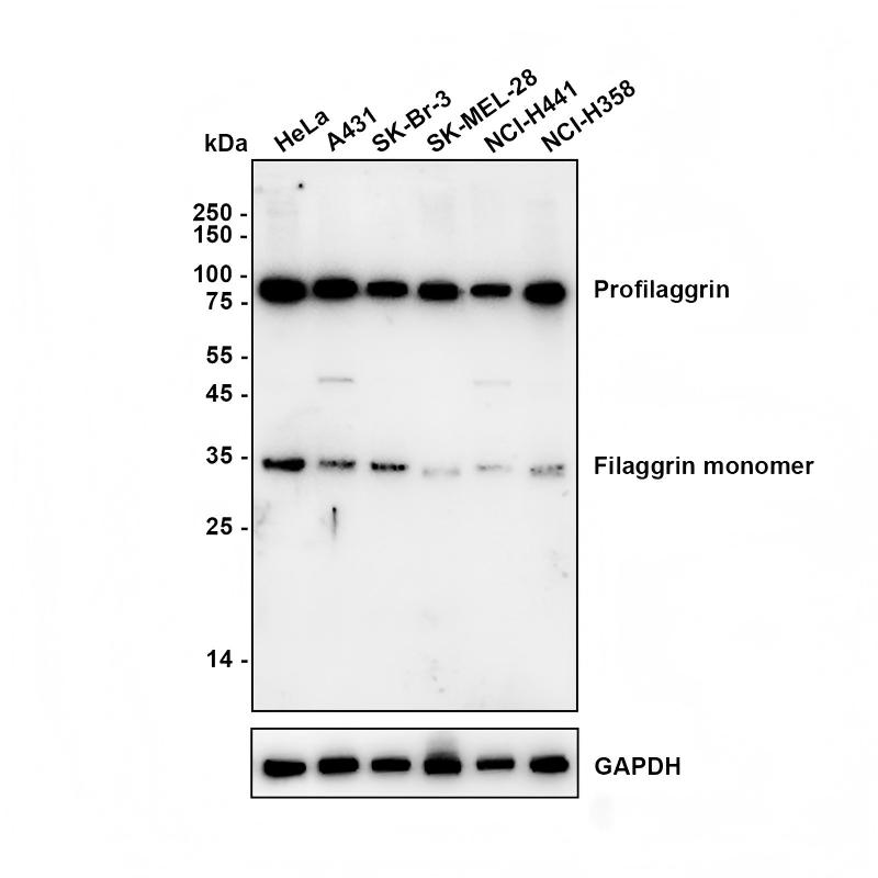
Western blot analysis of Filaggrin on different lysates with Rabbit anti-Filaggrin antibody at 1/2,000 dilution.Lysates/proteins at 15 µg/Lane. Exposure time: 3 minutes;Blocking: 5% NFDM/TBST, 1 hour at room temperature.Primary antibody:Rabbit anti-Filaggrin antibody, 1/2,000 in primary antibody dilution buffer,overnight at 4 °C.Secondary antibody: Goat anti-Rabbit IgG-HRP, 1/50,000 in 5% NFDM/TBST, 1 hour at room temperatureWB
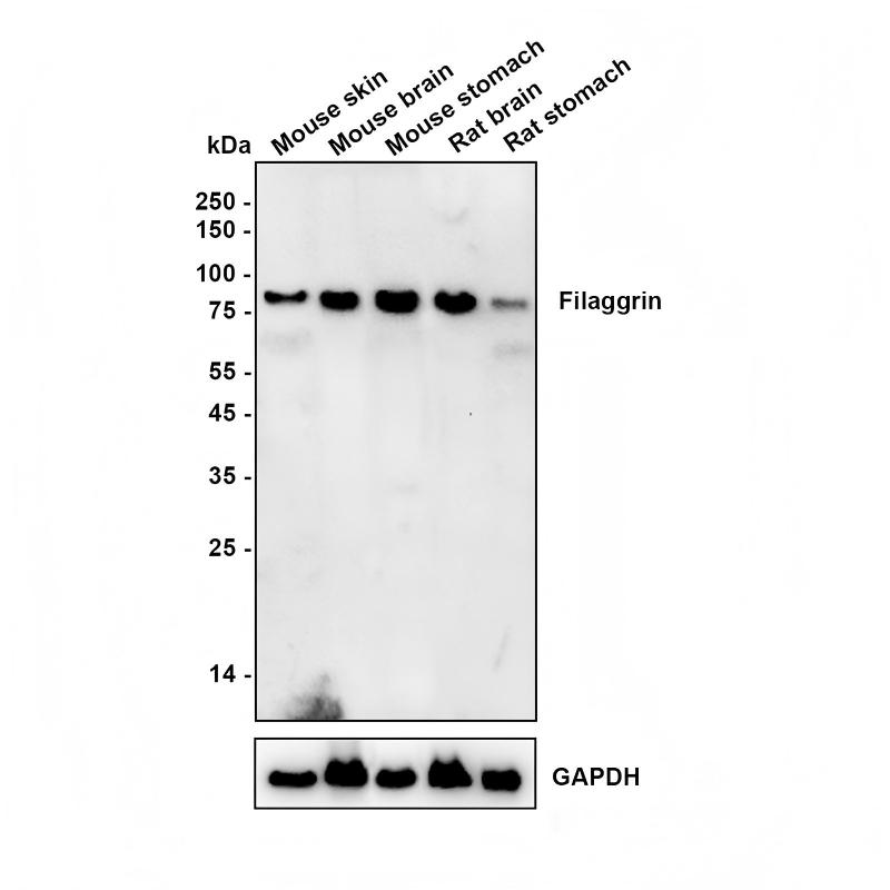
Western blot analysis of Filaggrin on different lysates with Rabbit anti-Filaggrin antibody at 1/5,000 dilution.ICC/IF
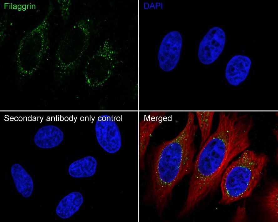
Application: Immunocytochemistry (IF-cell) Species: Human Sample: HeLa (Human cervix adenocarcinoma epithelial cell) Fixation: 4% Paraformaldehyde, 15 minutes at room temperature. Permeabilization: 0.1% Triton X-100, 15 minutes at room temperature. Blocking: 1% BSA + 10% normal goat serum, 1 hour at room temperature. Antibody dilution buffer: 1% BSA in PBST.Primary antibody:Rabbit anti-Filaggrin antibody, 1/50, overnight at 4°C. Secondary antibody: Goat Anti-Rabbit IgG (488), 45 minutes at room temperature.Counterstain: Beta tubulin (red), 1/100, overnight at 4℃. The Nuclear counterstain was DAPI (Blue).IHC
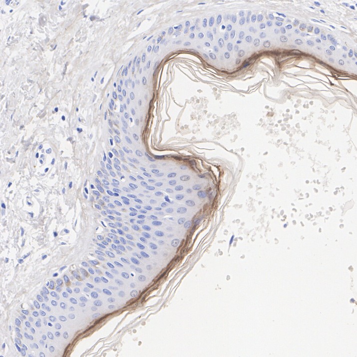
Application: Immunohistochemistry (IHC-P) Species: Human Tissue: Skin Sample: Paraffin-embedded section Antigen retrieval: Heat-mediated, Tris-EDTA buffer (pH 9.0), 20 minutes at 95°C. Wash buffer: 1× TBST Endogenous peroxidase blocking: 3% H₂O₂, 10 minutes at room temperature. Blocking: 1% BSA + 10% normal goat serum, 10 minutes at room temperature.Primary antibody:Rabbit anti-Filaggrin antibody.1/1,000, 1 hour at room temperature.Secondary antibody:Goat anti-Rabbit IgG (H&L) - HRP,20 minutes at room temperature.IHC
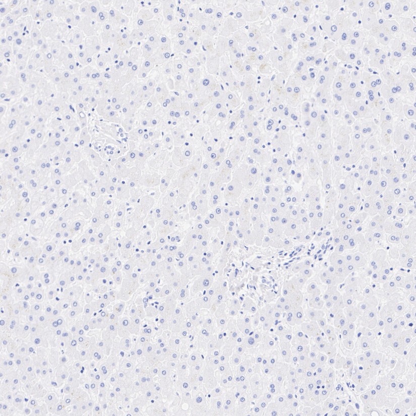
Application: Immunohistochemistry (IHC-P) Species: Human Tissue: Liver Sample: Paraffin-embedded section Antigen retrieval: Heat-mediated, Tris-EDTA buffer (pH 9.0), 20 minutes at 95°C. Wash buffer: 1× TBST Endogenous peroxidase blocking: 3% H₂O₂, 10 minutes at room temperature. Blocking: 1% BSA + 10% normal goat serum, 10 minutes at room temperature.Primary antibody: Rabbit anti-Filaggrin antibody, 1/1,000, 1 hour at room temperature.Secondary antibody:Goat anti-Rabbit IgG (H&L) - HRP, 20 minutes at room temperature. Negative expression of Filaggrin protein in liver is consistent with the predicted expression pattern.IF-P
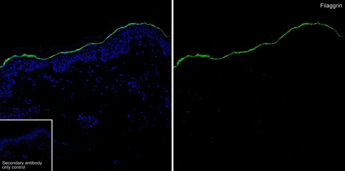
Application: Immunofluorescence (IF-tissue). Species: Human. Tissue: Skin. Sample: Paraffin-embedded section. Antigen retrieval: Heat-mediated, Tris-EDTA buffer (pH 9.0), 20 minutes at 95°C. Wash buffer: 1× PBS. Endogenous peroxidase blocking: 3% H₂O₂, 10 minutes.Blocking: 1% BSA + 10% normal goat serum, 10 minutes at room temperature. Primary antibody:Rabbit anti-Filaggrin antibody , 1/200, overnight at 4°C.Secondary antibody: Goat Anti-Rabbit IgG (488), 1.5 hours at room temperature.FC
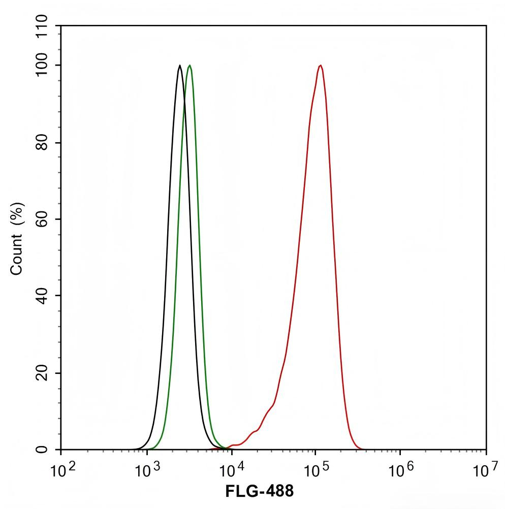
Application: Flow Cytometry (Intra) Species: Human Sample: HeLa (Human cervix adenocarcinoma epithelial cell) Fixation: 4% Paraformaldehyde, 15 minutes at room temperature. Permeabilization: 1x Permeabilization Buffer (eBioscience), 15 minutes at room temperature. Blocking: 1% BSA + 10% normal goat serum, 15 minutes at room temperature. Antibody dilution buffer: 1x PBS.Primary antibody:Rabbit anti-Filaggrin antibody (1/200) (Red) compared with Rabbit IgG Isotype Control( Green), 15 minutes at room temperature.Secondary antibody: Goat Anti-Rabbit IgG (488), 15 minutes at room temperature.| Product Name | Filaggrin Recombinant Rabbit Monoclonal Antibody |
|---|---|
| Antibody Type | Primary Antibodies |
| Immunogen | Recombinant protein within human Filaggrin aa 51-300. |
| Clonality | Monoclonal |
|---|---|
| Isotype | IgG |
| Host Species | Rabbit |
| Tested Applications | FCICC/IFIF-PIHCWB |
| WB:1:2000-1:5000 ICC/IF:1:50 IHC:1:1000 IF-P:1:200 FC:1:200 |
|
| Species Reactivity | HumanMouseRat |
| Concentration | 1mg/ml |
| Purification | Protein A |
| Gene Symbol | FLG |
|---|---|
| Gene Synonyms | FLG1 ATOD2 FLG-1 |
| Gene Full Name | filaggrin |
| Gene Summary | The protein encoded by this gene is an intermediate filament-associated protein that aggregates keratin intermediate filaments in mammalian epidermis. It is initially synthesized as a polyprotein precursor, profilaggrin (consisting of multiple filaggrin units of 324 aa each), which is localized in keratohyalin granules, and is subsequently proteolytically processed into individual functional filaggrin molecules. Mutations in this gene are associated with ichthyosis vulgaris.[provided by RefSeq, Dec 2009] |
| Alternative Names | Filaggrin FLG |
| Molecular Weight(MW) | 435 kDa(Observed band size: 37,77 kDa) |
| Cellular Localization | Cytoplasmic granule. |
WB
Western blot analysis of Filaggrin on different lysates with Rabbit anti-Filaggrin antibody at 1/2,000 dilution.Lysates/proteins at 15 µg/Lane. Exposure time: 3 minutes;Blocking: 5% NFDM/TBST, 1 hour at room temperature.Primary antibody:Rabbit anti-Filaggrin antibody, 1/2,000 in primary antibody dilution buffer,overnight at 4 °C.Secondary antibody: Goat anti-Rabbit IgG-HRP, 1/50,000 in 5% NFDM/TBST, 1 hour at room temperature
WB
Western blot analysis of Filaggrin on different lysates with Rabbit anti-Filaggrin antibody at 1/5,000 dilution.
ICC/IF
Application: Immunocytochemistry (IF-cell) Species: Human Sample: HeLa (Human cervix adenocarcinoma epithelial cell) Fixation: 4% Paraformaldehyde, 15 minutes at room temperature. Permeabilization: 0.1% Triton X-100, 15 minutes at room temperature. Blocking: 1% BSA + 10% normal goat serum, 1 hour at room temperature. Antibody dilution buffer: 1% BSA in PBST.Primary antibody:Rabbit anti-Filaggrin antibody, 1/50, overnight at 4°C. Secondary antibody: Goat Anti-Rabbit IgG (488), 45 minutes at room temperature.Counterstain: Beta tubulin (red), 1/100, overnight at 4℃. The Nuclear counterstain was DAPI (Blue).
IHC
Application: Immunohistochemistry (IHC-P) Species: Human Tissue: Skin Sample: Paraffin-embedded section Antigen retrieval: Heat-mediated, Tris-EDTA buffer (pH 9.0), 20 minutes at 95°C. Wash buffer: 1× TBST Endogenous peroxidase blocking: 3% H₂O₂, 10 minutes at room temperature. Blocking: 1% BSA + 10% normal goat serum, 10 minutes at room temperature.Primary antibody:Rabbit anti-Filaggrin antibody.1/1,000, 1 hour at room temperature.Secondary antibody:Goat anti-Rabbit IgG (H&L) - HRP,20 minutes at room temperature.
IHC
Application: Immunohistochemistry (IHC-P) Species: Human Tissue: Liver Sample: Paraffin-embedded section Antigen retrieval: Heat-mediated, Tris-EDTA buffer (pH 9.0), 20 minutes at 95°C. Wash buffer: 1× TBST Endogenous peroxidase blocking: 3% H₂O₂, 10 minutes at room temperature. Blocking: 1% BSA + 10% normal goat serum, 10 minutes at room temperature.Primary antibody: Rabbit anti-Filaggrin antibody, 1/1,000, 1 hour at room temperature.Secondary antibody:Goat anti-Rabbit IgG (H&L) - HRP, 20 minutes at room temperature. Negative expression of Filaggrin protein in liver is consistent with the predicted expression pattern.
IF-P
Application: Immunofluorescence (IF-tissue). Species: Human. Tissue: Skin. Sample: Paraffin-embedded section. Antigen retrieval: Heat-mediated, Tris-EDTA buffer (pH 9.0), 20 minutes at 95°C. Wash buffer: 1× PBS. Endogenous peroxidase blocking: 3% H₂O₂, 10 minutes.Blocking: 1% BSA + 10% normal goat serum, 10 minutes at room temperature. Primary antibody:Rabbit anti-Filaggrin antibody , 1/200, overnight at 4°C.Secondary antibody: Goat Anti-Rabbit IgG (488), 1.5 hours at room temperature.
FC
Application: Flow Cytometry (Intra) Species: Human Sample: HeLa (Human cervix adenocarcinoma epithelial cell) Fixation: 4% Paraformaldehyde, 15 minutes at room temperature. Permeabilization: 1x Permeabilization Buffer (eBioscience), 15 minutes at room temperature. Blocking: 1% BSA + 10% normal goat serum, 15 minutes at room temperature. Antibody dilution buffer: 1x PBS.Primary antibody:Rabbit anti-Filaggrin antibody (1/200) (Red) compared with Rabbit IgG Isotype Control( Green), 15 minutes at room temperature.Secondary antibody: Goat Anti-Rabbit IgG (488), 15 minutes at room temperature.| Application Notes | WB:1:2000-1:5000 ICC/IF:1:50 IHC:1:1000 IF-P:1:200 FC:1:200 |
|---|
| Form | Liquid |
|---|---|
| Storage Instructions | Shipped at 4℃. Store at +4℃ short term (1-2 weeks). It is recommended to aliquot into single-use upon delivery. Store at -20℃ long term. |
| Storage Buffer | PBS (pH7.4), 0.1% BSA, 40% Glycerol. Preservative: 0.05% Sodium Azide. |


