Application
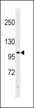
FGFR4 Antibody (E39) western blot analysis in 293 cell line lysates (35ug/lane). This demonstrates the FGFR4 antibody detected the FGFR4 protein (arrow).Application
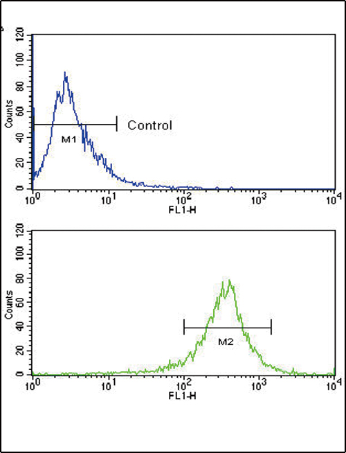
Flow cytometric analysis of WiDr cells using FGFR4 Antibody (N-term) (bottom histogram) compared to a negative control (top histogram). FITC-conjugated goat-anti-rabbit secondary antibodies were used for the analysis.Application
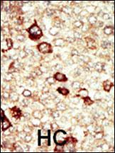
Formalin-fixed and paraffin-embedded human cancer tissue reacted with the primary FGFR4 Antibody (E39) , which was peroxidase-conjugated to the secondary antibody, followed by AEC staining. This data demonstrates the use of this antibody for immunohistochemistry; clinical relevance has not been evaluated. HC = hepatocarcinoma.Application
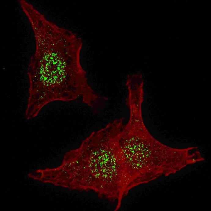
Fluorescent confocal image of HeLa cells stained with FGFR4 (N-term) antibody . HeLa cells were fixed with 4% PFA (20 min), permeabilized with Triton X-100 (0.2%, 30 min). Cells were then incubated with FGFR4 (N-term) primary antibody (1:200, 2 h at room temperature). For secondary antibody, Alexa Fluor® 488 conjugated donkey anti-rabbit antibody (green) was used (1:1000, 1h). Nuclei were counterstained with Hoechst 33342 (blue) (10 μg/ml, 5 min).| Product Name | FGFR4 antibody |
|---|---|
| Antibody Type | Primary Antibodies |
| Product description | Rabbit polyclonal to FGFR4Protein kinases are enzymes that transfer a phosphate group from a phosphate donor, generally the g phosphate of ATP, onto an acceptor amino acid in a substrate protein. By this basic mechanism, protein kinases mediate most of the signal transduction in eukaryotic cells, regulating cellular metabolism, transcription, cell cycle progression, cytoskeletal rearrangement and cell movement, apoptosis, and differentiation. With more than 500 gene products, the protein kinase family is one of the largest families of proteins in eukaryotes. The family has been classified in 8 major groups based on sequence comparison of their tyrosine (PTK) or serine/threonine (STK) kinase catalytic domains. The tyrosine kinase (TK) group is mainly involved in the regulation of cell-cell interactions such as differentiation, adhesion, motility and death. There are currently about 90 TK genes sequenced, 58 are of receptor protein TK (e.g. EGFR, EPH, FGFR, PDGFR, TRK, and VEGFR families), and 32 of cytosolic TK (e.g. ABL, FAK, JAK, and SRC families). |
| Immunogen | Synthetic peptide conjugated to KLH, corresponding to amino acids 39-56 of Human FGFR 4. |
| Clonality | Polyclonal |
|---|---|
| Isotype | IgG |
| Host Species | Rabbit |
| Tested Applications | FACSICC/IFIHC-PWB |
| For ELISA: Use at a dilution of 1:900 - 1:1,100. For IHC-F and IHC-P: Use at a dilution of 1:50 - 1:100. For WB: Use at a dilution of 1:100 - 1:500. Detects a band of approximately 96 kDa. Optimal dilutions/concentrations should be determined by the researcher. | |
| Species Reactivity | Human |
| Concentration | 0.2mg/ml |
| Purification | Protein G |
| Gene Synonyms | 2264, P22455, 134935, FGFR4, TKF, MGC20292, JTK2 |
|---|---|
| Target | FGFR4 |
| Cellular Localization | Plasma membrane |
Application
FGFR4 Antibody (E39) western blot analysis in 293 cell line lysates (35ug/lane). This demonstrates the FGFR4 antibody detected the FGFR4 protein (arrow).
Application
Flow cytometric analysis of WiDr cells using FGFR4 Antibody (N-term) (bottom histogram) compared to a negative control (top histogram). FITC-conjugated goat-anti-rabbit secondary antibodies were used for the analysis.
Application
Formalin-fixed and paraffin-embedded human cancer tissue reacted with the primary FGFR4 Antibody (E39) , which was peroxidase-conjugated to the secondary antibody, followed by AEC staining. This data demonstrates the use of this antibody for immunohistochemistry; clinical relevance has not been evaluated. HC = hepatocarcinoma.
Application
Fluorescent confocal image of HeLa cells stained with FGFR4 (N-term) antibody . HeLa cells were fixed with 4% PFA (20 min), permeabilized with Triton X-100 (0.2%, 30 min). Cells were then incubated with FGFR4 (N-term) primary antibody (1:200, 2 h at room temperature). For secondary antibody, Alexa Fluor® 488 conjugated donkey anti-rabbit antibody (green) was used (1:1000, 1h). Nuclei were counterstained with Hoechst 33342 (blue) (10 μg/ml, 5 min).| Positive Control | THP-1 cell lysate. |
|---|---|
| Application Notes | For ELISA: Use at a dilution of 1:900 - 1:1,100. For IHC-F and IHC-P: Use at a dilution of 1:50 - 1:100. For WB: Use at a dilution of 1:100 - 1:500. Detects a band of approximately 96 kDa. Optimal dilutions/concentrations should be determined by the researcher. |
| Form | Liquid |
|---|---|
| Storage Instructions | Keep as concentrated solution. Store at 4ºC short term. For extended storage aliquot and store at -20ºC or below. Avoid freeze-thaw cycles. |
| Storage Buffer | Phosphate buffered saline containing 0.09% sodium azide |


