Application
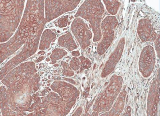
Immunohistochemical analysis of paraffin-embedded Oral CA Cal27 xenograft, using FGFRL1 antibody at 1:100 dilution.Application
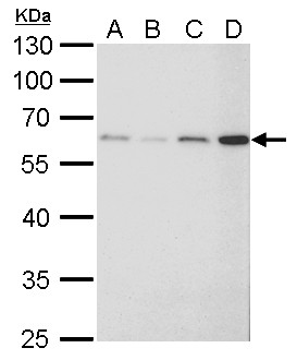
FGFR-5 antibody detects FGFR-5 protein by western blot analysis. A. 30 µg 293T whole cell lysate/extract B. 30 µg A431 whole cell lysate/extract C. 30 µg HeLa whole cell lysate/extract D. 30 µg HepG2 whole cell lysate/extract 10% SDS-PAGE FGFR-5 antibody dilution: 1:1000 The HRP-conjugated anti-rabbit IgG antibody was used to detect the primary antibody.Application
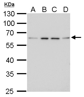
FGFR-5 antibody detects FGFR-5 protein by western blot analysis. A. 30 µg Jurkat whole cell lysate/extract B. 30 µg Raji whole cell lysate/extract C. 30 µg K562 whole cell lysate/extract D. 30 µg NCI-H929 whole cell lysate/extract 10% SDS-PAGE FGFR-5 antibody dilution: 1:1000 The HRP-conjugated anti-rabbit IgG antibody was used to detect the primary antibody.Application
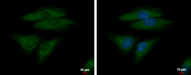
FGFR-5 antibody detects FGFR-5 protein at cytoplasm by immunofluorescent analysis. Sample: HeLa cells were fixed in 4% paraformaldehyde at RT for 15 min. Green: FGFR-5 protein stained by FGFR-5 antibody diluted at 1:500. Blue: Hoechst 33342 staining. Scale bar = 10 μm.| Product Name | FGFR-5 antibody |
|---|---|
| Antibody Type | Primary Antibodies |
| Product description | Rabbit polyclonal antibody to FGFR-5 (fibroblast growth factor receptor-like 1)The protein encoded by this gene is a member of the fibroblast growth factor receptor (FGFR) family, where amino acid sequence is highly conserved between members and throughout evolution. FGFR family members differ from one another in their ligand affinities and tissue distribution. A full-length representative protein would consist of an extracellular region, composed of three immunoglobulin-like domains, a single hydrophobic membrane-spanning segment and a cytoplasmic tyrosine kinase domain. The extracellular portion of the protein interacts with fibroblast growth factors, setting in motion a cascade of downstream signals, ultimately influencing mitogenesis and differentiation. A marked difference between this gene product and the other family members is its lack of a cytoplasmic tyrosine kinase domain. The result is a transmembrane receptor that could interact with other family members and potentially inhibit signaling. Multiple alternatively spliced transcript variants encoding the same isoform have been found for this gene. [provided by RefSeq] |
| Immunogen | Recombinant protein encompassing a sequence within the center region of human FGFR-5. The exact sequence is proprietary. |
| Clonality | Polyclonal |
|---|---|
| Isotype | IgG |
| Host Species | Rabbit |
| Tested Applications | ICC/IFIHC-PWB |
| *Optimal dilutions/concentrations should be determined by the researcher. | |
| Species Reactivity | Human |
| Predicted Reactivity | Mouse, Rat, BovineMouse (86%), Rat (86%), Bovine (89%) |
| Concentration | 1mg/ml (Please refer to the vial label for the specific concentration) |
| Purification | Affinity purified |
| Gene Synonyms | FGFR5 antibody, FHFR antibody, FGFRL1 antibody, FGFR-like protein antibody, FGF homologous factor receptor antibody, FGFR-5 antibody, FGF receptor-like protein 1 antibody, fibroblast growth factor receptor-like 1 antibody, fibroblast growth factor receptor |
|---|---|
| Target | FGFR-5 |
| Molecular Weight(MW) | 55 kDa (note) (3) |
| Cellular Localization | Membrane; Single-pass type I membrane protein |
Application
Immunohistochemical analysis of paraffin-embedded Oral CA Cal27 xenograft, using FGFRL1 antibody at 1:100 dilution.
Application
FGFR-5 antibody detects FGFR-5 protein by western blot analysis. A. 30 µg 293T whole cell lysate/extract B. 30 µg A431 whole cell lysate/extract C. 30 µg HeLa whole cell lysate/extract D. 30 µg HepG2 whole cell lysate/extract 10% SDS-PAGE FGFR-5 antibody dilution: 1:1000 The HRP-conjugated anti-rabbit IgG antibody was used to detect the primary antibody.
Application
FGFR-5 antibody detects FGFR-5 protein by western blot analysis. A. 30 µg Jurkat whole cell lysate/extract B. 30 µg Raji whole cell lysate/extract C. 30 µg K562 whole cell lysate/extract D. 30 µg NCI-H929 whole cell lysate/extract 10% SDS-PAGE FGFR-5 antibody dilution: 1:1000 The HRP-conjugated anti-rabbit IgG antibody was used to detect the primary antibody.
Application
FGFR-5 antibody detects FGFR-5 protein at cytoplasm by immunofluorescent analysis. Sample: HeLa cells were fixed in 4% paraformaldehyde at RT for 15 min. Green: FGFR-5 protein stained by FGFR-5 antibody diluted at 1:500. Blue: Hoechst 33342 staining. Scale bar = 10 μm.| Positive Control | 293T , A431 , HeLa , Jurkat , Raji , K562 , NCI-H929 , HepG2 , *MCF7 |
|---|---|
| Application Notes | *Optimal dilutions/concentrations should be determined by the researcher. |
| Form | Liquid |
|---|---|
| Storage Instructions | Keep as concentrated solution. Aliquot and store at -20ºC or below. Avoid multiple freeze-thaw cycles. |
| Storage Buffer | PBS |


