Application
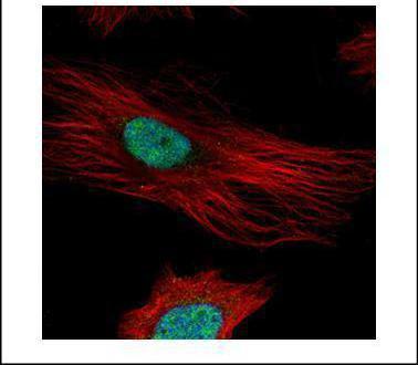
Confocal immunofluorescence analysis (Olympus FV10i) of paraformaldehyde-fixed HeLa, using Fibrillarin antibody (Green) at 1:500 dilution. Alpha-tubulin filaments were labeled with (Red) at 1:2000.Application
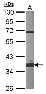
Sample (30 ug of whole cell lysate) A:NIH-3T3 10% SDS PAGE diluted at 1:1000Application
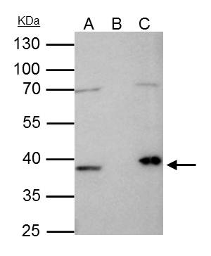
Fibrillarin antibody immunoprecipitates FBL protein in IP experiments. IP samples: HeLa nuclear extract A. 35 μg HeLa nuclear extract B. Control with 4 μg of preimmune Rabbit IgG C. Immunoprecipitation of FBL protein by 4 μg Fibrillarin antibody 10 % SDS-PAGE The immunoprecipitated FBL protein was detected by Fibrillarin antibody diluted at 1:1000. [EasyBlot anti-rabbit IgG was used as a secondary reagent]Application
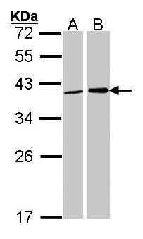
Sample (30 ug of whole cell lysate) A: 293T B: H1299 12% SDS PAGE diluted at 1:1000Application
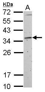
Fibrillarin antibody detects FBL protein by Western blot analysis. A. 30 µg Rat2 whole cell lysate/extract 12 % SDS-PAGE Fibrillarin antibody dilution: 1:1000| Product Name | Fibrillarin antibody |
|---|---|
| Antibody Type | Primary Antibodies |
| Product description | Rabbit Polyclonal antibody to Fibrillarin (fibrillarin)This gene product is a component of a nucleolar small nuclear ribonucleoprotein (snRNP) particle thought to participate in the first step in processing preribosomal RNA. It is associated with the U3, U8, and U13 small nuclear RNAs and is located in the dense fibrillar component (DFC) of the nucleolus. The encoded protein contains an N-terminal repetitive domain that is rich in glycine and arginine residues, like fibrillarins in other species. Its central region resembles an RNA-binding domain and contains an RNP consensus sequence. Antisera from approximately 8% of humans with the autoimmune disease scleroderma recognize fibrillarin. [provided by RefSeq] |
| Immunogen | Recombinant protein encompassing a sequence within the center region of human Fibrillarin. The exact sequence is proprietary. |
| Clonality | Polyclonal |
|---|---|
| Isotype | IgG |
| Host Species | Rabbit |
| Tested Applications | ICC/IFIPWB |
| *Optimal dilutions/concentrations should be determined by the researcher.
: |
|
| Species Reactivity | HumanMouseRat |
| Predicted Reactivity | Zebrafish, BovineZebrafish (80%), Bovine (98%) |
| Concentration | 1mg/ml (Please refer to the vial label for the specific concentration) |
| Purification | Affinity purified |
| Gene Synonyms | FIB antibody FLRN antibody RNU3IP1 antibody FBL antibody 34 kDa nucleolar scleroderma antigen antibody RNA U3 small nucleolar interacting protein 1 antibody rRNA 2'-O-methyltransferase fibrillarin antibody 34-kD nucleolar scleroderma antigen antibo |
|---|---|
| Molecular Weight(MW) | 34 kDa (note) |
| Cellular Localization | Nucleus , nucleolus |
Application
Confocal immunofluorescence analysis (Olympus FV10i) of paraformaldehyde-fixed HeLa, using Fibrillarin antibody (Green) at 1:500 dilution. Alpha-tubulin filaments were labeled with (Red) at 1:2000.
Application
Sample (30 ug of whole cell lysate) A:NIH-3T3 10% SDS PAGE diluted at 1:1000
Application
Fibrillarin antibody immunoprecipitates FBL protein in IP experiments. IP samples: HeLa nuclear extract A. 35 μg HeLa nuclear extract B. Control with 4 μg of preimmune Rabbit IgG C. Immunoprecipitation of FBL protein by 4 μg Fibrillarin antibody 10 % SDS-PAGE The immunoprecipitated FBL protein was detected by Fibrillarin antibody diluted at 1:1000. [EasyBlot anti-rabbit IgG was used as a secondary reagent]
Application
Sample (30 ug of whole cell lysate) A: 293T B: H1299 12% SDS PAGE diluted at 1:1000
Application
Fibrillarin antibody detects FBL protein by Western blot analysis. A. 30 µg Rat2 whole cell lysate/extract 12 % SDS-PAGE Fibrillarin antibody dilution: 1:1000| Positive Control | 293T , H1299 , NIH-3T3 , Rat2 |
|---|---|
| Application Notes | *Optimal dilutions/concentrations should be determined by the researcher.
: |
| Form | Liquid |
|---|---|
| Storage Instructions | Keep as concentrated solution. Aliquot and store at -20ºC or below. Avoid multiple freeze-thaw cycles. |
| Storage Buffer | 1XPBS, 40% Glycerol (pH7). 0.01% Thimerosal was added as a preservative. |


