Application
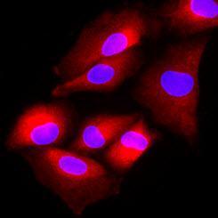
Immunofluorescence of human A549 cells stained with Hoechst 33342 (Blue) and monoclonal anti-human FUS2 antibody (1:500) with Texas Red (red).Application
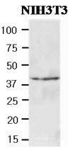
Cell lysates of NIH3T3(40ug) were resolved by SDS-PAGE, transferred to NC membrane and probed with anti-human FUS2 (1:500). Proteins were visualized using a goat anti-mouse secondary antibody conjugated to HRP and an ECL detection system.| Product Name | FUS2 antibody |
|---|---|
| Antibody Type | Primary Antibodies |
| Product description | Mouse monoclonal antibody [AT2F4] to FUS2Vertebrate FUS2 genes, which are known to be putative tumor suppressor gene, contain several important domains such as the catalytic N-acetyltransferase (NAT) domain. NAT domain is essential enzymes involved in several sophisticated cellular processes such as N-acetylation, O-acetylatin. NAT enzymes may be involved in susceptibility to cancer including colorectal cancer because of the presence of carcinogenic heterocyclic amines in some cooked foods. FUS2 was physically localized to the cytoplasm. Also, FUS2 showed the actin dependent movement, closely related to the polarization in the budding yeast, Saccharomyces cerevisiae. |
| Immunogen | Recombinant FUS2 (1-308aa) purified from E. coli |
| Clonality | Monoclonal |
|---|---|
| Isotype | IgG1 |
| Clone Number | AT2F4 |
| Host Species | Mouse |
| Tested Applications | ICC/IFWB |
| ICC/IF:1:200~1:500 WB:1:500~1:2000 Notes:Optimal dilutions/concentrations should be determined by the researcher. |
|
| Species Reactivity | Human |
| Concentration | 1mg/ml (Please refer to the vial label for the specific concentration) |
| Purification | Affinity purified |
| Gene Synonyms | N acetyltransferase6 Protein fusion2. Protein fusion 2. Protein fus2 N acetyltransferase 6 Protein fus 2 FUS-2 Protein fusion-2 FUS2 FUS 2 Protein fusion2 NAT6 Protein fusion 2 |
|---|---|
| Target | FUS2 |
| Light Chain Type | kappa |
Application
Immunofluorescence of human A549 cells stained with Hoechst 33342 (Blue) and monoclonal anti-human FUS2 antibody (1:500) with Texas Red (red).
Application
Cell lysates of NIH3T3(40ug) were resolved by SDS-PAGE, transferred to NC membrane and probed with anti-human FUS2 (1:500). Proteins were visualized using a goat anti-mouse secondary antibody conjugated to HRP and an ECL detection system.| Application Notes | ICC/IF:1:200~1:500 WB:1:500~1:2000 Notes:Optimal dilutions/concentrations should be determined by the researcher. |
|---|
| Form | Liquid |
|---|---|
| Storage Instructions | Can be stored at +4C. For long term storage, aliquot and store at -20C. Avoid repeated freezing and thawing cycles. |
| Storage Buffer | In Phosphate-Buffered Saline (pH 7.4) with 0.1% Sodium Azide |


