Application
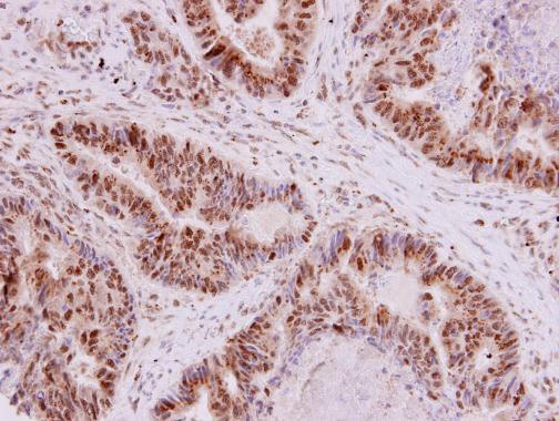
G9a antibody detects G9a protein at nucleus in human rectal carcinoma by immunohistochemical analysis. Sample: Paraffin-embedded human rectal carcinoma. G9a antibody diluted at 1:250.Application
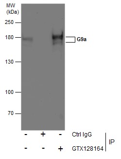
Immunoprecipitation of G9a protein from HeLa whole cell extracts using 5 μg of G9a antibody . Western blot analysis was performed using G9a antibody . EasyBlot anti-Rabbit IgG was used as a secondary reagent.Application
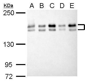
G9a antibody detects EHMT2 protein by western blot analysis. A. 30 µg 293T whole cell lysate/extract B. 30 µg A431 whole cell lysate/extract C. 30 µg HeLa whole cell lysate/extract D. 30 µg HepG2 whole cell lysate/extract E. 30 µg A375 whole cell lysate/extract 5% SDS-PAGE G9a antibody dilution: 1:1000 The HRP-conjugated anti-rabbit IgG antibody was used to detect the primary antibody.Application
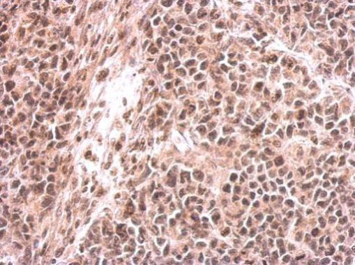
G9a antibody detects EHMT2 protein at on HeLa xenograft by immunohistochemical analysis. Sample: Paraffin-embedded HeLa xenograft. G9a antibody dilution: 1:500.Application
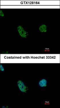
G9a antibody detects EHMT2 protein at cytoplasm by immunofluorescent analysis. Sample: HeLa cells were fixed in 4% paraformaldehyde at RT for 5 min. Green: EHMT2 protein stained by G9a antibody diluted at 1:500. Blue: Hoechst 33342 staining.Application
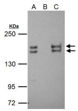
EHMT2 antibody immunoprecipitates EHMT2 protein in IP experiments. IP Sample: 2 mg HeLa whole cell lysate/extract A. 40 μg HeLa whole cell lysate/extract B. Control with 2 μg of preimmune rabbit IgG C. Immunoprecipitation of EHMT2 protein by 2 μg of EHMT2 antibody 5% SDS-PAGE The immunoprecipitated EHMT2 protein was detected by EHMT2 antibody diluted at 1:1000. EasyBlot anti-rabbit IgG was used as a secondary reagent.Application
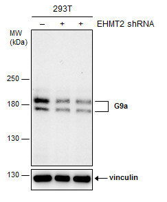
Non-transfected (–) and transfected (+) 293T whole cell extracts (30 µg) were separated by 5% SDS-PAGE, and the membrane was blotted with G9a antibody diluted at 1:1000. The HRP-conjugated anti-rabbit IgG antibody was used to detect the primary antibody.| Product Name | G9a antibody |
|---|---|
| Antibody Type | Primary Antibodies |
| Product description | Rabbit Polyclonal antibody to G9a (euchromatic histone-lysine N-methyltransferase 2)A cluster of genes, BAT1-BAT5, has been localized in the vicinity of the genes for TNF alpha and TNF beta. This gene is found near this cluster; it was mapped near the gene for C2 within a 120-kb region that included a HSP70 gene pair. These genes are all within the human major histocompatibility complex class III region. This gene was thought to be two different genes, NG36 and G9a, adjacent to each other but a recent publication shows that there is only a single gene. The protein encoded by this gene is thought to be involved in intracellular protein-protein interaction. There are three alternatively spliced transcript variants of this gene but only two are fully described. [provided by RefSeq] |
| Immunogen | Carrier-protein conjugated synthetic peptide encompassing a sequence within the N-terminus region of human G9a. The exact sequence is proprietary. |
| Clonality | Polyclonal |
|---|---|
| Isotype | IgG |
| Host Species | Rabbit |
| Tested Applications | ICC/IFIHC-PIPWB |
| *Optimal dilutions/concentrations should be determined by the researcher.
: |
|
| Species Reactivity | Human |
| Predicted Reactivity | Rat, BovineRat (100%), Bovine (100%) |
| Concentration | 1mg/ml (Please refer to the vial label for the specific concentration) |
| Purification | Affinity purified |
| Gene Synonyms | BAT8 antibody C6orf30 antibody DKFZp686H08213 antibody FLJ35547 antibody G9A antibody KMT1C antibody NG36 antibody EHMT2 antibody protein G9a antibody histone-lysine N-methyltransferase H3 lysine-9 specific 3 antibody ankyrin repeat-containing p |
|---|---|
| Molecular Weight(MW) | 132 kDa (note) (3) |
| Cellular Localization | Nucleus |
Application
G9a antibody detects G9a protein at nucleus in human rectal carcinoma by immunohistochemical analysis. Sample: Paraffin-embedded human rectal carcinoma. G9a antibody diluted at 1:250.
Application
Immunoprecipitation of G9a protein from HeLa whole cell extracts using 5 μg of G9a antibody . Western blot analysis was performed using G9a antibody . EasyBlot anti-Rabbit IgG was used as a secondary reagent.
Application
G9a antibody detects EHMT2 protein by western blot analysis. A. 30 µg 293T whole cell lysate/extract B. 30 µg A431 whole cell lysate/extract C. 30 µg HeLa whole cell lysate/extract D. 30 µg HepG2 whole cell lysate/extract E. 30 µg A375 whole cell lysate/extract 5% SDS-PAGE G9a antibody dilution: 1:1000 The HRP-conjugated anti-rabbit IgG antibody was used to detect the primary antibody.
Application
G9a antibody detects EHMT2 protein at on HeLa xenograft by immunohistochemical analysis. Sample: Paraffin-embedded HeLa xenograft. G9a antibody dilution: 1:500.
Application
G9a antibody detects EHMT2 protein at cytoplasm by immunofluorescent analysis. Sample: HeLa cells were fixed in 4% paraformaldehyde at RT for 5 min. Green: EHMT2 protein stained by G9a antibody diluted at 1:500. Blue: Hoechst 33342 staining.
Application
EHMT2 antibody immunoprecipitates EHMT2 protein in IP experiments. IP Sample: 2 mg HeLa whole cell lysate/extract A. 40 μg HeLa whole cell lysate/extract B. Control with 2 μg of preimmune rabbit IgG C. Immunoprecipitation of EHMT2 protein by 2 μg of EHMT2 antibody 5% SDS-PAGE The immunoprecipitated EHMT2 protein was detected by EHMT2 antibody diluted at 1:1000. EasyBlot anti-rabbit IgG was used as a secondary reagent.
Application
Non-transfected (–) and transfected (+) 293T whole cell extracts (30 µg) were separated by 5% SDS-PAGE, and the membrane was blotted with G9a antibody diluted at 1:1000. The HRP-conjugated anti-rabbit IgG antibody was used to detect the primary antibody.| Positive Control | 293T , A431 , HeLa , HepG2 , A375 |
|---|---|
| Application Notes | *Optimal dilutions/concentrations should be determined by the researcher.
: |
| Form | Liquid |
|---|---|
| Storage Instructions | Keep as concentrated solution. Aliquot and store at -20ºC or below. Avoid multiple freeze-thaw cycles. |
| Storage Buffer | 1XPBS, 20% Glycerol (pH7). 0.01% Thimerosal was added as a preservative. |


