Application
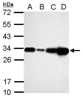
Sample (30 ug of whole cell lysate) A: A549 B: H1299 C: HCT116 D: MCF-7 12% SDS PAGE diluted at 1:10000Application
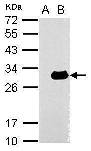
Sample (30 ug of whole cell lysate) A: Non-transfected 293T lysates B: LGALS3 transfected 293T lysates 12% SDS PAGE diluted at 1:10000Application
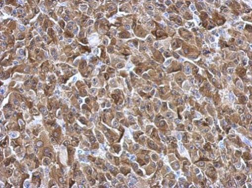
Immunohistochemical analysis of paraffin-embedded HBL435 xenograft, using Galectin3 antibody at 1:500 dilution.Application
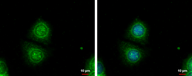
Galectin3 antibody detects Galectin3 protein at cytoplasm by immunofluorescent analysis. Sample: MCF-7 cells were fixed in ice-cold MeOH for 5 min. Green: Galectin3 protein stained by Galectin3 antibody diluted at 1:1000. Blue: Hoechst 33342 staining. Scale bar = 10 μm.Application
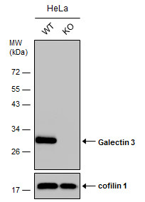
Wild-type (WT) and Galectin 3 knownout (KO) HeLa cell extracts (30 µg) were separated by 12% SDS-PAGE, and the membrane was blotted with Galectin 3 antibody diluted at 1:10000. The HRP-conjugated anti-rabbit IgG antibody was used to detect the primary antibody.Application
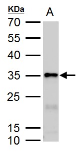
Galectin3 antibody detects Galectin3 protein by western blot analysis. A. 50 μg rat colon lysate/extract 12 % SDS-PAGE Galectin3 antibody dilution: 1:10000| Product Name | Galectin 3 antibody |
|---|---|
| Antibody Type | Primary Antibodies |
| Product description | Rabbit Polyclonal antibody to Galectin 3 (galectin-3)This gene encodes a member of the galectin family of carbohydrate binding proteins. Members of this protein family have an affinity for beta-galactosides. The encoded protein is characterized by an N-terminal proline-rich tandem repeat domain and a single C-terminal carbohydrate recognition domain. This protein can self-associate through the N-terminal domain allowing it to bind to multivalent saccharide ligands. This protein localizes to the extracellular matrix, the cytoplasm and the nucleus. This protein plays a role in numerous cellular functions including apoptosis, innate immunity, cell adhesion and T-cell regulation. Alternate splicing results in multiple transcript variants. |
| Immunogen | Recombinant protein encompassing a sequence within the center region of human Galectin3. The exact sequence is proprietary. |
| Clonality | Polyclonal |
|---|---|
| Isotype | IgG |
| Host Species | Rabbit |
| Tested Applications | ICC/IFIHC-PWB |
| *Optimal dilutions/concentrations should be determined by the researcher.
: |
|
| Species Reactivity | HumanRat |
| Predicted Reactivity | Mouse, Dog, Pig, Rhesus MonkeyMouse (81%), Dog (84%), Pig (81%), Rhesus Monkey (91%) |
| Concentration | 1mg/ml (Please refer to the vial label for the specific concentration) |
| Purification | Affinity purified |
| Gene Synonyms | CBP35 antibody GAL3 antibody GALBP antibody GALIG antibody L31 antibody LGALS2 antibody MAC2 antibody LGALS3 antibody IgE-binding protein antibody carbohydrate-binding protein 35 antibody 35 kDa lectin antibody MAC-2 antigen antibody galactose- |
|---|---|
| Molecular Weight(MW) | 26 kDa (note) (3) |
| Cellular Localization | Nucleus , Cytoplasm , Secreted |
Application
Sample (30 ug of whole cell lysate) A: A549 B: H1299 C: HCT116 D: MCF-7 12% SDS PAGE diluted at 1:10000
Application
Sample (30 ug of whole cell lysate) A: Non-transfected 293T lysates B: LGALS3 transfected 293T lysates 12% SDS PAGE diluted at 1:10000
Application
Immunohistochemical analysis of paraffin-embedded HBL435 xenograft, using Galectin3 antibody at 1:500 dilution.
Application
Galectin3 antibody detects Galectin3 protein at cytoplasm by immunofluorescent analysis. Sample: MCF-7 cells were fixed in ice-cold MeOH for 5 min. Green: Galectin3 protein stained by Galectin3 antibody diluted at 1:1000. Blue: Hoechst 33342 staining. Scale bar = 10 μm.
Application
Wild-type (WT) and Galectin 3 knownout (KO) HeLa cell extracts (30 µg) were separated by 12% SDS-PAGE, and the membrane was blotted with Galectin 3 antibody diluted at 1:10000. The HRP-conjugated anti-rabbit IgG antibody was used to detect the primary antibody.
Application
Galectin3 antibody detects Galectin3 protein by western blot analysis. A. 50 μg rat colon lysate/extract 12 % SDS-PAGE Galectin3 antibody dilution: 1:10000| Positive Control | A549 , H1299 , HCT116 , MCF-7 , Recombinant LGALS3 protein , rat colon |
|---|---|
| Application Notes | *Optimal dilutions/concentrations should be determined by the researcher.
: |
| Form | Liquid |
|---|---|
| Storage Instructions | Keep as concentrated solution. Aliquot and store at -20ºC or below. Avoid multiple freeze-thaw cycles. |
| Storage Buffer | 1XPBS, 20% Glycerol (pH7). 0.01% Thimerosal was added as a preservative. |


