Application
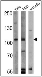
Western blot analysis of GAP in 25 ug of HeLa, A431 and mouse brain lysates. Proteins were transferred to a PVDF membrane and blocked with a blocking buffer at 4?C overnight. The membrane was probed with GAP antibody [B4F8] at a dilution of 1:200 overnight at 4°C, washed in TBST, and probed with an HRP-conjugated secondary antibody for 1 hr at room temperature in the dark. Chemiluminescent detection was performed.Application
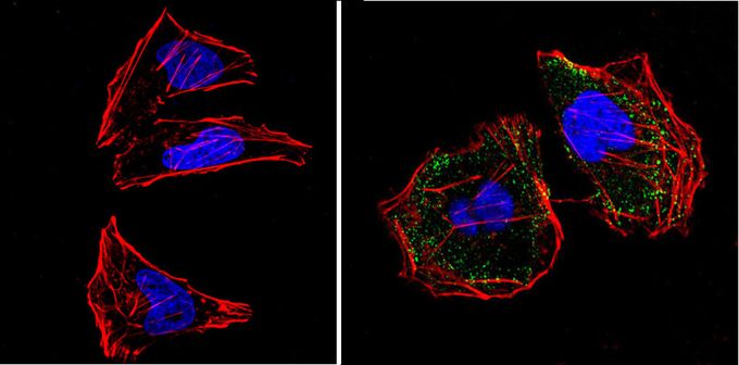
Immunofluorescent analysis of GAP (green) in HeLa cells (right) compared with a negative control in the absence of primary antibody (left). Formalin-fixed cells were permeabilized with 0.1% Triton X-100 in TBS for 5-10 minutes, blocked with 3% BSA-PBS for 30 minutes at room temperature and probed with GAP antibody [B4F8] in 3% BSA-PBS at a dilution of 1:200 and incubated overnight at 4?C in a humidified chamber. Cells were washed with PBST and incubated with a proper secondary antibody. F-actin (red) was stained with a flourescent red phalloidin and nuclei (blue) were stained with DAPI for 5-10 minutes in the dark. Images were taken at a magnification of 60x.| Product Name | GAP antibody |
|---|---|
| Antibody Type | Primary Antibodies |
| Product description | Mouse monoclonal antibody [B4F8] to GAPThe protein encoded by this gene is located in the cytoplasm and is part of the GAP1 family of GTPase-activating proteins. The gene product stimulates the GTPase activity of normal RAS p21 but not its oncogenic counterpart. Acting as a suppressor of RAS function, the protein enhances the weak intrinsic GTPase activity of RAS proteins resulting in the inactive GDP-bound form of RAS, thereby allowing control of cellular proliferation and differentiation. Mutations leading to changes in the binding sites of either protein are associated with basal cell carcinomas. Mutations also have been associated with hereditary capillary malformations (CM) with or without arteriovenous malformations (AVM) and Parkes Weber syndrome. Alternative splicing results in two isoforms where the shorter isoform, lacking the N-terminal hydrophobic region but retaining the same activity, appears to be abundantly expressed in placental but not adult tissues. [provided by RefSeq, May 2012] |
| Immunogen | Full length recombinant human GAP. |
| Clonality | Monoclonal |
|---|---|
| Isotype | IgG2a |
| Clone Number | B4F8 |
| Host Species | Mouse |
| Tested Applications | ICC/IFIPWB |
| ICC/IF: 5 µg/ml: |
|
| Species Reactivity | HumanMouseRatPrimateBovine |
| Concentration | 1mg/ml |
| Purification | Protein A |
| Gene Synonyms | p120RASGAP CMAVM RASA1 RASGAP RASA PKWS GAP CM-AVM RAS p21 protein activator (GTPase activating protein) 1 p120GAP |
|---|---|
| Target | GTPase Activating Protein |
Application
Western blot analysis of GAP in 25 ug of HeLa, A431 and mouse brain lysates. Proteins were transferred to a PVDF membrane and blocked with a blocking buffer at 4?C overnight. The membrane was probed with GAP antibody [B4F8] at a dilution of 1:200 overnight at 4°C, washed in TBST, and probed with an HRP-conjugated secondary antibody for 1 hr at room temperature in the dark. Chemiluminescent detection was performed.
Application
Immunofluorescent analysis of GAP (green) in HeLa cells (right) compared with a negative control in the absence of primary antibody (left). Formalin-fixed cells were permeabilized with 0.1% Triton X-100 in TBS for 5-10 minutes, blocked with 3% BSA-PBS for 30 minutes at room temperature and probed with GAP antibody [B4F8] in 3% BSA-PBS at a dilution of 1:200 and incubated overnight at 4?C in a humidified chamber. Cells were washed with PBST and incubated with a proper secondary antibody. F-actin (red) was stained with a flourescent red phalloidin and nuclei (blue) were stained with DAPI for 5-10 minutes in the dark. Images were taken at a magnification of 60x.| Application Notes | ICC/IF: 5 µg/ml: |
|---|
| Form | Liquid |
|---|---|
| Storage Instructions | -20° C, Avoid Freeze/Thaw Cycles |
| Storage Buffer | phosphate buffered saline , pH 7.4, 150mM NaCl, 0.02% sodium azide and 50% glycerol. |


