Application
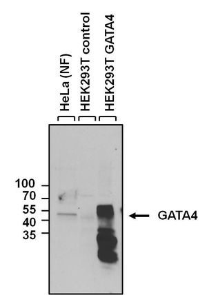
WB analysis of HeLa nuclear fraction (NF), HEK293T control and GATA4 over-expressing HEK293T lysates using GATA4 antibody at a dilution of 1:1000.Application
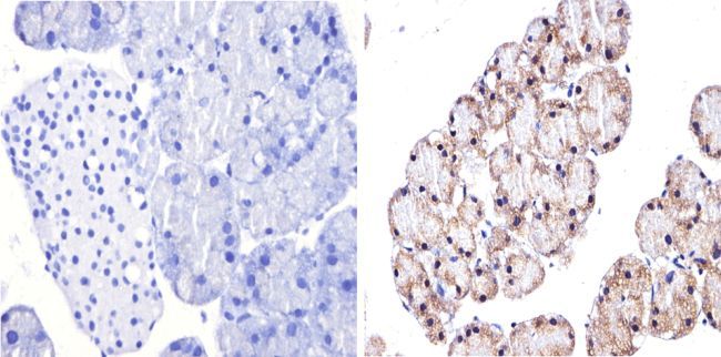
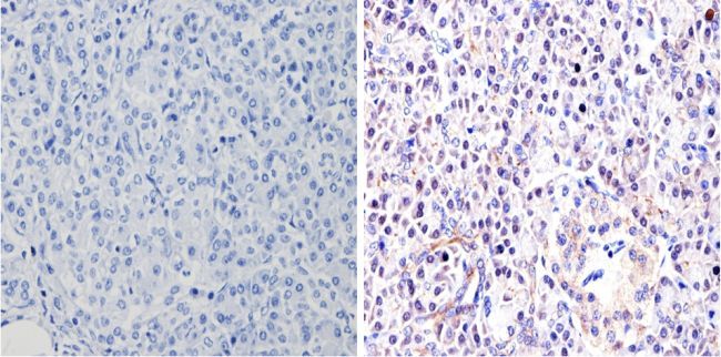
IHC-P analysis of mouse pancreas tissues with (right) or without (left) GATA4 antibody at a dilution of 1:500. To expose target proteins, antigen retrieval method was performed using 10mM sodium citrate (pH 6.0) microwaved for 8-15 min.Application
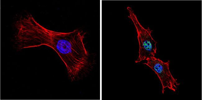
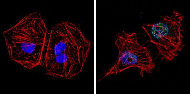
ICC/IF analysis of NIH-3T3 cells with (right) or without (left) GATA4 antibody at a dilution of 1:100 (green). F-actin (red) was stained with a flourescent red phalloidin and nuclei (blue) were stained with DAPI. Images were taken at a magnification of 60x.Application
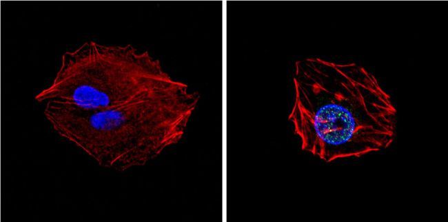
ICC/IF analysis of HepG2 cells with (right) or without (left) GATA4 antibody at a dilution of 1:200 (green). F-actin (red) was stained with a flourescent red phalloidin and nuclei (blue) were stained with DAPI. Images were taken at a magnification of 60x.Application
ICC/IF analysis of HeLa cells with (right) or without (left) GATA4 antibody at a dilution of 1:200 (green). F-actin (red) was stained with a flourescent red phalloidin and nuclei (blue) were stained with DAPI. Images were taken at a magnification of 60x.Application
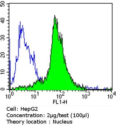
FACS analysis of HepG2 cells using GATA4 antibody (green) or an isotype control (blue).Application
IHC-P analysis of human pancreas tissues with (right) or without (left) GATA4 antibody at a dilution of 1:200. To expose target proteins, antigen retrieval method was performed using 10mM sodium citrate (pH 6.0) microwaved for 8-15 min.Application
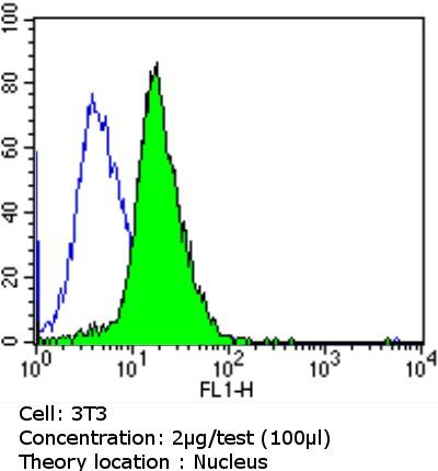
FACS analysis of NIH/3T3 cells using GATA4 antibody (green) or an isotype control (blue).Application
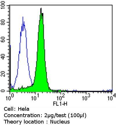
FACS analysis of HeLa cells using GATA4 antibody (green) or an isotype control (blue).Application
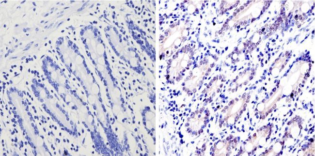
IHC-P analysis of human small intestine tissues with (right) or without (left) GATA4 antibody. To expose target proteins, antigen retrieval method was performed using 10mM sodium citrate (pH 6.0) microwaved for 8-15 min.| Product Name | GATA4 antibody |
|---|---|
| Antibody Type | Primary Antibodies |
| Product description | Rabbit polyclonal antibody to GATA4This gene encodes a member of the GATA family of zinc-finger transcription factors. Members of this family recognize the GATA motif which is present in the promoters of many genes. This protein is thought to regulate genes involved in embryogenesis and in myocardial differentiation and function. Mutations in this gene have been associated with cardiac septal defects. [provided by RefSeq, Jul 2008] |
| Immunogen | Pooled peptides: RKEGIQTRKRKPKNLNKSK & CKQDSWNSLVLADSHGDIITA |
| Clonality | Polyclonal |
|---|---|
| Isotype | IgG |
| Host Species | Rabbit |
| Tested Applications | FACSICC/IFIHC-PWB |
| Species Reactivity | HumanMouse |
| Concentration | 1mg/ml |
| Purification | Affinity purified |
| Gene Synonyms | VSD1 , GATA binding protein 4 , ASD2 |
|---|---|
| Target | GATA4 |
Application
WB analysis of HeLa nuclear fraction (NF), HEK293T control and GATA4 over-expressing HEK293T lysates using GATA4 antibody at a dilution of 1:1000.
Application
IHC-P analysis of mouse pancreas tissues with (right) or without (left) GATA4 antibody at a dilution of 1:500. To expose target proteins, antigen retrieval method was performed using 10mM sodium citrate (pH 6.0) microwaved for 8-15 min.
Application
ICC/IF analysis of NIH-3T3 cells with (right) or without (left) GATA4 antibody at a dilution of 1:100 (green). F-actin (red) was stained with a flourescent red phalloidin and nuclei (blue) were stained with DAPI. Images were taken at a magnification of 60x.
Application
ICC/IF analysis of HepG2 cells with (right) or without (left) GATA4 antibody at a dilution of 1:200 (green). F-actin (red) was stained with a flourescent red phalloidin and nuclei (blue) were stained with DAPI. Images were taken at a magnification of 60x.
Application
ICC/IF analysis of HeLa cells with (right) or without (left) GATA4 antibody at a dilution of 1:200 (green). F-actin (red) was stained with a flourescent red phalloidin and nuclei (blue) were stained with DAPI. Images were taken at a magnification of 60x.
Application
FACS analysis of HepG2 cells using GATA4 antibody (green) or an isotype control (blue).
Application
IHC-P analysis of human pancreas tissues with (right) or without (left) GATA4 antibody at a dilution of 1:200. To expose target proteins, antigen retrieval method was performed using 10mM sodium citrate (pH 6.0) microwaved for 8-15 min.
Application
FACS analysis of NIH/3T3 cells using GATA4 antibody (green) or an isotype control (blue).
Application
FACS analysis of HeLa cells using GATA4 antibody (green) or an isotype control (blue).
Application
IHC-P analysis of human small intestine tissues with (right) or without (left) GATA4 antibody. To expose target proteins, antigen retrieval method was performed using 10mM sodium citrate (pH 6.0) microwaved for 8-15 min.| Form | Liquid |
|---|---|
| Storage Instructions | -20° C |
| Storage Buffer | PBS with 1mg/ml BSA, 30% glycerol |


