Application
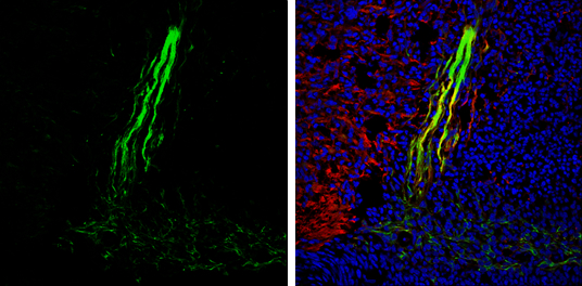
Gephyrin antibody [N2C1], Internal detects Gephyrin protein expression by immunohistochemical analysis. Sample: Frozen sectioned E13.5 Rat brain. Green: Gephyrin protein stained by Gephyrin antibody [N2C1], Internal diluted at 1:250. Red: beta Tubulin 3/ TUJ1, a mature neuron marker, stained by beta Tubulin 3/ TUJ1 antibody [GT11710] diluted at 1:500. Blue: Fluoroshield with DAPI .Application
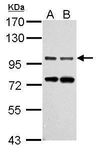
Sample (30 ug of whole cell lysate) A: NIH-3T3 B: JC 7.5% SDS PAGE diluted at 1:1000Application
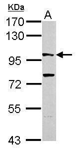
Sample (30 ug of whole cell lysate) A: PC-12 7.5% SDS PAGE diluted at 1:1000Application
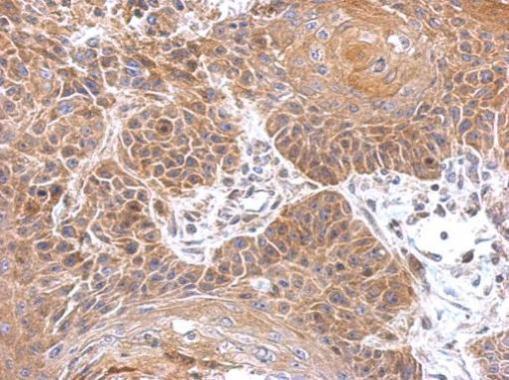
Immunohistochemical analysis of paraffin-embedded mouse brain, using Gephyrin antibody at 1:500 dilution.Application
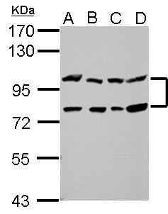
Sample (30 ug of whole cell lysate) A: 293T B: A431 C: HeLa D: HepG2 7.5% SDS PAGE diluted at 1:1000Application
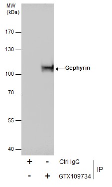
Immunoprecipitation of Gephyrin protein from A431 whole cell extracts using 5 μg of Gephyrin antibody [N2C1], Internal . Western blot analysis was performed using Gephyrin antibody [N2C1], Internal . EasyBlot anti-Rabbit IgG was used as a secondary reagent.| Product Name | Gephyrin antibody , Internal |
|---|---|
| Antibody Type | Primary Antibodies |
| Product description | Rabbit Polyclonal antibody to Gephyrin (gephyrin)This gene encodes a neuronal assembly protein that anchors inhibitory neurotransmitter receptors to the postsynaptic cytoskeleton via high affinity binding to a receptor subunit domain and tubulin dimers. In nonneuronal tissues, the encoded protein is also required for molybdenum cofactor biosynthesis. Mutations in this gene may be associated with the neurological condition hyperplexia and also lead to molybdenum cofactor deficiency. Numerous alternatively spliced transcript variants encoding different isoforms have been described; however, the full-length nature of all transcript variants is not currently known. [provided by RefSeq] |
| Immunogen | Recombinant protein encompassing a sequence within the center region of human Gephyrin. The exact sequence is proprietary. |
| Clonality | Polyclonal |
|---|---|
| Isotype | IgG |
| Host Species | Rabbit |
| Tested Applications | IHC-FIHC-PIPWB |
| *Optimal dilutions/concentrations should be determined by the researcher.
: |
|
| Species Reactivity | HumanMouseRat |
| Predicted Reactivity | Xenopus laevis, Pig, Chicken, Rhesus MonkeyXenopus laevis (99%), Pig (100%), Chicken (100%), Rhesus Monkey (100%) |
| Concentration | 1mg/ml (Please refer to the vial label for the specific concentration) |
| Purification | Affinity purified |
| Gene Synonyms | GEPH antibody GPH antibody GPHRYN antibody KIAA1385 antibody GPHN antibody gephyrin antibody |
|---|---|
| Molecular Weight(MW) | 80 kDa (note) (2) |
| Cellular Localization | Cell junction , synapse (By similarity) , synapse , postsynaptic cell membrane; Peripheral membrane protein; Cytoplasmic side (By similarity) , Cytoplasm , cytoskeleton (By similarity) |
Application
Gephyrin antibody [N2C1], Internal detects Gephyrin protein expression by immunohistochemical analysis. Sample: Frozen sectioned E13.5 Rat brain. Green: Gephyrin protein stained by Gephyrin antibody [N2C1], Internal diluted at 1:250. Red: beta Tubulin 3/ TUJ1, a mature neuron marker, stained by beta Tubulin 3/ TUJ1 antibody [GT11710] diluted at 1:500. Blue: Fluoroshield with DAPI .
Application
Sample (30 ug of whole cell lysate) A: NIH-3T3 B: JC 7.5% SDS PAGE diluted at 1:1000
Application
Sample (30 ug of whole cell lysate) A: PC-12 7.5% SDS PAGE diluted at 1:1000
Application
Immunohistochemical analysis of paraffin-embedded mouse brain, using Gephyrin antibody at 1:500 dilution.
Application
Sample (30 ug of whole cell lysate) A: 293T B: A431 C: HeLa D: HepG2 7.5% SDS PAGE diluted at 1:1000
Application
Immunoprecipitation of Gephyrin protein from A431 whole cell extracts using 5 μg of Gephyrin antibody [N2C1], Internal . Western blot analysis was performed using Gephyrin antibody [N2C1], Internal . EasyBlot anti-Rabbit IgG was used as a secondary reagent.| Positive Control | 293T , A431 , HeLa , HepG2 , NIH-3T3 , JC , PC-12 |
|---|---|
| Application Notes | *Optimal dilutions/concentrations should be determined by the researcher.
: |
| Form | Liquid |
|---|---|
| Storage Instructions | Keep as concentrated solution. Aliquot and store at -20ºC or below. Avoid multiple freeze-thaw cycles. |
| Storage Buffer | 1XPBS, 20% Glycerol (pH7). 0.01% Thimerosal was added as a preservative. |


