
Application
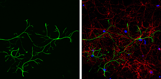
GFAP antibody detects GFAP protein expression in astrocytes and Tuj1 antibody detects Tuj1 protein expression in neurons by immunofluorescent analysis. Sample:Cultured rat E18 primary cortical neurons and astrocytes, DIV 8. Cells were fixed in 4% paraformaldehyde at RT for 15 min. Green: GFAP protein stained by GFAP antibody diluted at 1:250. Red: beta Tubulin 3/ TUJ1, stained by beta Tubulin 3/ TUJ1 antibody [GT11710] diluted at 1:250. Blue: Fluoroshield with DAPI .Application
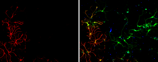
GFAP antibody detects GFAP protein expression by immunofluorescent analysis. Sample:Cultured rat E18 primary cortical neuron, DIV 8 cells were fixed in 4% paraformaldehyde at RT for 15 min. Red: GFAP protein stained by GFAP antibody diluted at 1:250. Green: Glutamine synthetase, stained by Glutamine synthetase antibody [GT7711] diluted at 1:250. Blue: Fluoroshield with DAPI .Application
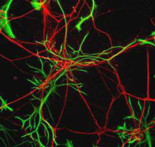
Immunocytochemistry/Immunofluorescence: GFAP Antibody - Rat neurons stained with Neurofilament Heavy antibody (red) and GFAP antibody (green).Application
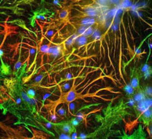
Immunocytochemistry/Immunofluorescence: GFAP Antibody - Mixed neuron-glial cultures stained with GFAP antibody (red) and Vimentin antibody (green). The fibroblastic cells contain only Vimentin and so are green. The astrocytes contain either Vimentin and GFAP (appearing golden) or predominantly GFAP (appearing red). Blue is nuclear DNA stain.Application
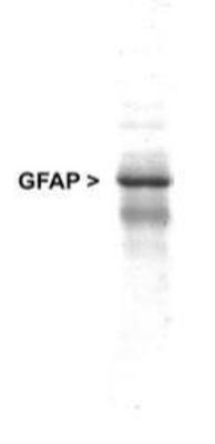
Western Blot: GFAP Antibody - Western blot analysis of GFAP expression in whole rat cerebellum homogenate usingWB
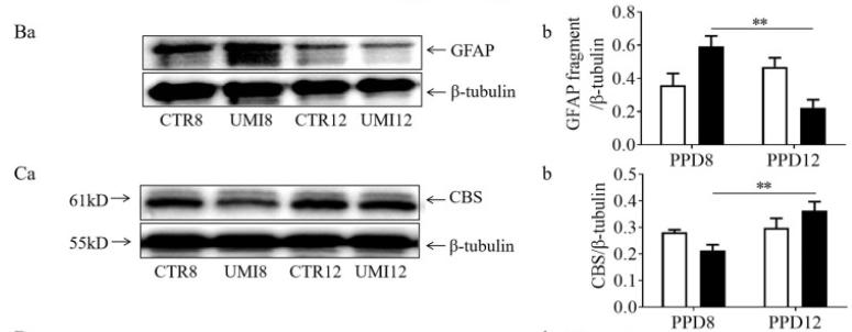
The summary graphs (b) showing GFAP filament/fiber length and fluorescence intensity of CBS-positive cells in ventral glial lamina (VGL, c). (B–C) Representative Western blot bands (a) and the summary graphs (b) showing GFAP fragments (B, n = 5) and CBS (C, n = 7), respectively.WB
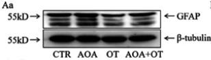
(A) Western blot bands (a) and the summary graphWB
(b) showing the expression of GFAP in the SON of brain slices (n = 6) with CTR, AOA, OT, and AOA plus OT, respectively.| Product Name | GFAP antibody |
|---|---|
| Antibody Type | Primary Antibodies |
| Product description | Rabbit polyclonal antibody [R40] to GFAPGlial fibrillary acidic protein (GFAP) is a member of the class III intermediate filament protein family. It is heavily and specifically expressed in astrocytes and certain other astroglia in the central nervous system, in satellite cells in peripheral ganglia, and in non-myelinating Schwann cells in peripheral nerves. In addition neural stem cells frequently strongly express GFAP. Antibodies to GFAP are therefore very useful as markers of astrocytic cells. In addition many types of brain tumor, presumably derived from astrocytic cells, heavily express GFAP. |
| Immunogen | Native GFAP purified from Pig Spinal Cord. |
| Conjugation | Agarose |
| Modification | Acetylated |
| Clonality | Polyclonal |
|---|---|
| Isotype | Cocktail |
| Host Species | Rabbit |
| Tested Applications | ICC/IFIHC-FIHC-PIPWB |
| Can be used to identify astrocytic cells, their progenitors and their descendents in tissue sections and in tissue culture. For WB: Use at a dilution of 1:5,000. A 55 kDa band can be seen in Western blotting and another at about 48kDa, apparently a breakdown product of the 55kDa band. For ICC/IF: 1:100-1:1000. Optimal dilutions/concentrations should be determined by the researcher.: |
|
| Species Reactivity | HumanMouseRat |
| Predicted Reactivity | Dog,Pig,Bovine |
| Concentration | 1mg/ml |
| Purification | Affinity purified |
| Gene Synonyms | P14136 396562 137780 FLJ45472 2670 GFAP |
|---|---|
| Target | GFAP |
| Tissue Specificity | Specifically recognizes mammalian GFAP on western blots and immunocytochemically. |
| Cellular Localization | Neural stem cell marker Astrocyte marker |
Application
GFAP antibody detects GFAP protein expression in astrocytes and Tuj1 antibody detects Tuj1 protein expression in neurons by immunofluorescent analysis. Sample:Cultured rat E18 primary cortical neurons and astrocytes, DIV 8. Cells were fixed in 4% paraformaldehyde at RT for 15 min. Green: GFAP protein stained by GFAP antibody diluted at 1:250. Red: beta Tubulin 3/ TUJ1, stained by beta Tubulin 3/ TUJ1 antibody [GT11710] diluted at 1:250. Blue: Fluoroshield with DAPI .
Application
GFAP antibody detects GFAP protein expression by immunofluorescent analysis. Sample:Cultured rat E18 primary cortical neuron, DIV 8 cells were fixed in 4% paraformaldehyde at RT for 15 min. Red: GFAP protein stained by GFAP antibody diluted at 1:250. Green: Glutamine synthetase, stained by Glutamine synthetase antibody [GT7711] diluted at 1:250. Blue: Fluoroshield with DAPI .
Application
Immunocytochemistry/Immunofluorescence: GFAP Antibody - Rat neurons stained with Neurofilament Heavy antibody (red) and GFAP antibody (green).
Application
Immunocytochemistry/Immunofluorescence: GFAP Antibody - Mixed neuron-glial cultures stained with GFAP antibody (red) and Vimentin antibody (green). The fibroblastic cells contain only Vimentin and so are green. The astrocytes contain either Vimentin and GFAP (appearing golden) or predominantly GFAP (appearing red). Blue is nuclear DNA stain.
Application
Western Blot: GFAP Antibody - Western blot analysis of GFAP expression in whole rat cerebellum homogenate using
WB
The summary graphs (b) showing GFAP filament/fiber length and fluorescence intensity of CBS-positive cells in ventral glial lamina (VGL, c). (B–C) Representative Western blot bands (a) and the summary graphs (b) showing GFAP fragments (B, n = 5) and CBS (C, n = 7), respectively.
WB
(A) Western blot bands (a) and the summary graph
WB
(b) showing the expression of GFAP in the SON of brain slices (n = 6) with CTR, AOA, OT, and AOA plus OT, respectively.| Application Notes | Can be used to identify astrocytic cells, their progenitors and their descendents in tissue sections and in tissue culture. For WB: Use at a dilution of 1:5,000. A 55 kDa band can be seen in Western blotting and another at about 48kDa, apparently a breakdown product of the 55kDa band. For ICC/IF: 1:100-1:1000. Optimal dilutions/concentrations should be determined by the researcher.: |
|---|
| Form | Liquid |
|---|---|
| Storage Instructions | Keep as concentrated solution. Store at 4ºC short term. For extended storage aliquot and store at -20ºC or below. Avoid freeze-thaw cycles. |
| Storage Buffer | serum with 0.01% sodium azide |


