Application
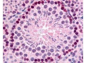
GeneTex's Affinity Purified anti-mouse Gli-2 antibody was used at 10 µg/ml to evaluate staining on several mouse tissues. Moderate to strong staining was seen on many tissues, with low background staining. This image shows Gli-2 staining of mouse testis. Tissue was formalin-fixed and paraffin embedded.Application
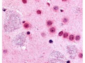
GeneTex's Affinity Purified anti-mouse Gli-2 antibody was used at 10 µg/ml to evaluate staining on several mouse tissues. Moderate to strong staining was seen on many tissues with low background staining. This image shows Gli-2 staining of mouse brain. Tissue was formalin-fixed and paraffin embedded.Application
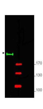
Western blot using GeneTex's Affinity Purified anti-Gli-2 antibody shows detection of a predominant band at ~190 kDa corresponding to Gli-2 (arrowhead) in mouse brain whole cell lysate (lane 1). Pre-incubation of antibody with immunizing peptide completely blocks staining of this band (lane 2). ~ 25 µg of lysate was resolved on a 4-8% Tris-glycine gel by SDS-PAGE and transferred onto nitrocellulose. After blocking with 5% goat serum and 0.5% BLOTTO in PBS, the membrane was probed with the primary antibody diluted to 1:750. Incubation was at room temperature for 2 h followed by washes and reaction with a 1:10,000 dilution of IRDye® 800 conjugated Gt-a-Rabbit IgG (H&L) MX10 for 45 min at room temperature. Molecular weight markers are shown (M) using the 700 nm channel (red). IRDye® 800 fluorescence image was captured using the Odyssey® Infrared Imaging System developed by LI-COR. IRDye is a trademark of LI-COR, Inc. Other detection systems will yield similar results.| Product Name | Gli2 antibody |
|---|---|
| Antibody Type | Primary Antibodies |
| Product description | Rabbit Polyclonal antibody to Gli2This protein belongs to the C2H2-type zinc finger protein subclass of the Gli family. Members of this subclass are characterized as transcription factors that bind DNA through zinc finger motifs. These motifs contain conserved H-C links. Gli family zinc finger proteins are mediators of Sonic hedgehog (Shh) signaling and they are implicated as potent oncogenes in the embryonal carcinoma cell. The protein encoded by this gene localizes to the cytoplasm and activates patched Drosophila homolog (PTCH) gene expression. Gli2 is also thought to play a role during embryogenesis. The encoded protein is associated with several phenotypes- Greig cephalopolysyndactyly syndrome, Pallister-Hall syndrome, preaxial polydactyly type IV, postaxial polydactyly types A1 and B. |
| Immunogen | Synthetic peptide: LRGAPSASRGSYTQQPR-C conjugated to KLH, corresponding to amino acids 1193-1209 of Mouse Gli2 |
| Clonality | Polyclonal |
|---|---|
| Isotype | IgG |
| Host Species | Rabbit |
| Tested Applications | ELISAIHCIHC-PWB |
| *Optimal dilutions/concentrations should be determined by the researcher.
: |
|
| Species Reactivity | HumanMouse |
| Concentration | 1mg/ml |
| Purification | Affinity purified |
| Gene Synonyms | AW546128 |
|---|---|
| Target | Gli2 |
Application
GeneTex's Affinity Purified anti-mouse Gli-2 antibody was used at 10 µg/ml to evaluate staining on several mouse tissues. Moderate to strong staining was seen on many tissues, with low background staining. This image shows Gli-2 staining of mouse testis. Tissue was formalin-fixed and paraffin embedded.
Application
GeneTex's Affinity Purified anti-mouse Gli-2 antibody was used at 10 µg/ml to evaluate staining on several mouse tissues. Moderate to strong staining was seen on many tissues with low background staining. This image shows Gli-2 staining of mouse brain. Tissue was formalin-fixed and paraffin embedded.
Application
Western blot using GeneTex's Affinity Purified anti-Gli-2 antibody shows detection of a predominant band at ~190 kDa corresponding to Gli-2 (arrowhead) in mouse brain whole cell lysate (lane 1). Pre-incubation of antibody with immunizing peptide completely blocks staining of this band (lane 2). ~ 25 µg of lysate was resolved on a 4-8% Tris-glycine gel by SDS-PAGE and transferred onto nitrocellulose. After blocking with 5% goat serum and 0.5% BLOTTO in PBS, the membrane was probed with the primary antibody diluted to 1:750. Incubation was at room temperature for 2 h followed by washes and reaction with a 1:10,000 dilution of IRDye® 800 conjugated Gt-a-Rabbit IgG (H&L) MX10 for 45 min at room temperature. Molecular weight markers are shown (M) using the 700 nm channel (red). IRDye® 800 fluorescence image was captured using the Odyssey® Infrared Imaging System developed by LI-COR. IRDye is a trademark of LI-COR, Inc. Other detection systems will yield similar results.| Application Notes | *Optimal dilutions/concentrations should be determined by the researcher.
: |
|---|
| Form | Liquid |
|---|---|
| Storage Instructions | Store vial at -20° C prior to opening. Aliquot contents and freeze at -20° C or below for extended storage. Avoid cycles of freezing and thawing. |
| Storage Buffer | 0.02 M Potassium Phosphate, 0.15 M Sodium Chloride, pH 7.2 and 0.01% (w/v) Sodium Azide |


