Application
Immunofluorescence analysis of paraformaldehyde-fixed A549, using Glucokinase antibody at 1:200 dilution.Application
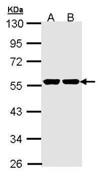
Sample (30 µg of whole cell lysate) A: H1299 B: HeLa 10% SDS PAGE diluted at 1:1000 The HRP-conjugated anti-rabbit IgG antibody was used to detect the primary antibody.Application
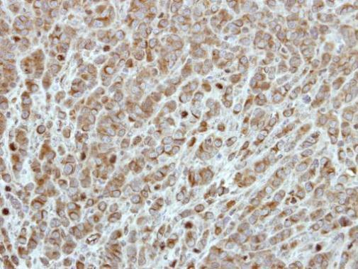
Immunohistochemical analysis of paraffin-embedded MCF7 xenograft, using Glucokinase antibody at 1:500 dilution.Application
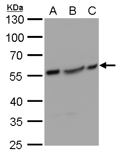
GCK antibody detects GCK protein by western blot analysis. A. 30 µg Huh7 whole cell lysate/extract B. 30 µg Hep3B whole cell lysate/extract C. 30 µg HepG2 whole cell lysate/extract 10% SDS-PAGE GCK antibody dilution: 1:1000 The HRP-conjugated anti-rabbit IgG antibody was used to detect the primary antibody.Application
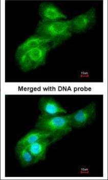
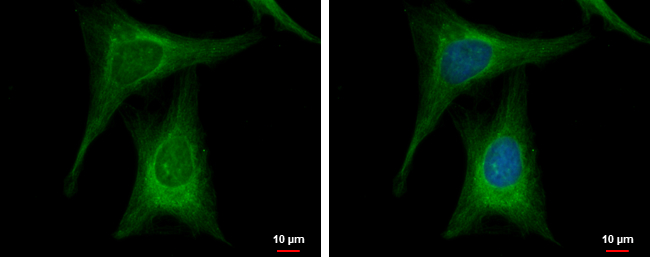
Glucokinase antibody [N1C1-2] detects Glucokinase protein at cytoplasm by immunofluorescent analysis. Sample: HeLa cells were fixed in 4% paraformaldehyde at RT for 15 min. Green: Glucokinase protein stained by Glucokinase antibody [N1C1-2] diluted at 1:500. Blue: Hoechst 33342 staining.| Product Name | Glucokinase antibody |
|---|---|
| Antibody Type | Primary Antibodies |
| Product description | Rabbit Polyclonal antibody to Glucokinase (glucokinase (hexokinase 4))Hexokinases phosphorylate glucose to produce glucose-6-phosphate, thus committing glucose to the glycolytic pathway. Alternative splicing of this gene results in three tissue-specific forms of glucokinase, one found in pancreatic islet beta cells and two found in liver. The protein localizes to the outer membrane of mitochondria. In contrast to other forms of hexokinase, this enzyme is not inhibited by its product glucose-6-phosphate but remains active while glucose is abundant. Mutations in this gene have been associated with non-insulin dependent diabetes mellitus (NIDDM), maturity onset diabetes of the young, type 2 (MODY2) and persistent hyperinsulinemic hypoglycemia of infancy (PHHI). [provided by RefSeq] |
| Immunogen | Recombinant protein encompassing a sequence within the center region of human Glucokinase. The exact sequence is proprietary. |
| Modification | Acetylated |
| Clonality | Polyclonal |
|---|---|
| Isotype | IgG |
| Host Species | Rabbit |
| Tested Applications | ICC/IFIHC-PWB |
| *Optimal dilutions/concentrations should be determined by the researcher.: |
|
| Species Reactivity | HumanMonkeyMouseRat |
| Predicted Reactivity | Mouse, Rat, Zebrafish, Xenopus laevis, Cat, Sheep, BovineMouse (97%), Rat (97%), Zebrafish (86%), Xenopus laevis (87%), Cat (96%), Sheep (97%), Bovine (96%) |
| Concentration | 1mg/ml (Please refer to the vial label for the specific concentration) |
| Purification | Affinity purified |
| Gene Synonyms | FGQTL3 antibody GK antibody GLK antibody HHF3 antibody HK4 antibody HKIV antibody HXKP antibody LGLK antibody MODY2 antibody GCK antibody hexokinase-4 antibody ATP:D-hexose 6-phosphotransferase antibody hexokinase type IV antibody hexokinase-D |
|---|---|
| Molecular Weight(MW) | 52 kDa (note) (3) |
Application
Immunofluorescence analysis of paraformaldehyde-fixed A549, using Glucokinase antibody at 1:200 dilution.
Application
Sample (30 µg of whole cell lysate) A: H1299 B: HeLa 10% SDS PAGE diluted at 1:1000 The HRP-conjugated anti-rabbit IgG antibody was used to detect the primary antibody.
Application
Immunohistochemical analysis of paraffin-embedded MCF7 xenograft, using Glucokinase antibody at 1:500 dilution.
Application
GCK antibody detects GCK protein by western blot analysis. A. 30 µg Huh7 whole cell lysate/extract B. 30 µg Hep3B whole cell lysate/extract C. 30 µg HepG2 whole cell lysate/extract 10% SDS-PAGE GCK antibody dilution: 1:1000 The HRP-conjugated anti-rabbit IgG antibody was used to detect the primary antibody.
Application
Glucokinase antibody [N1C1-2] detects Glucokinase protein at cytoplasm by immunofluorescent analysis. Sample: HeLa cells were fixed in 4% paraformaldehyde at RT for 15 min. Green: Glucokinase protein stained by Glucokinase antibody [N1C1-2] diluted at 1:500. Blue: Hoechst 33342 staining.| Positive Control | 293T , A431 , H1299 , HeLa , HepG2 , Molt-4 , Raji , Huh7 , Hep3B , HepG2 |
|---|---|
| Application Notes | *Optimal dilutions/concentrations should be determined by the researcher.: |
| Form | Liquid |
|---|---|
| Storage Instructions | Keep as concentrated solution. Aliquot and store at -20ºC or below. Avoid multiple freeze-thaw cycles. |
| Storage Buffer | 0.1M Tris, 0.1M Glycine, 10% Glycerol (pH7). 0.01% Thimerosal was added as a preservative. |


