Application
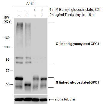
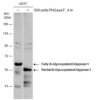
Untreated (–) and treated (+) A431 whole cell extracts (30 µg) were separated by 7.5% SDS-PAGE, and the membrane was blotted with Glypican 1 antibody [N3C3] diluted at 1:3000. The HRP-conjugated anti-rabbit IgG antibody was used to detect the primary antibody.Application
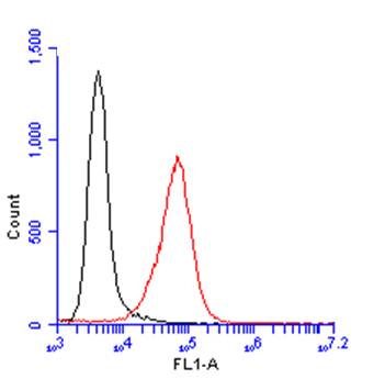
Glypican 1 antibody [N3C3] detects Glypican 1 protein by flow cytometry analysis. Sample: A431 cell. Black: Unlabelled sample was used as a control. Red: Glypican 1 antibody [N3C3] dilution: 1:100. Acquisition of 20,000 events were collected using a Dylight 488-conjugated secondary antibody for FACS analysis.Application
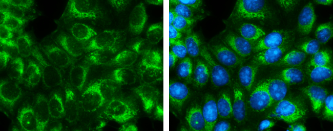
Glypican 1 antibody [N3C3] detects Glypican 1 protein at endoplasmic reticulum by immunofluorescent analysis. Sample: MCF7 cells were fixed in ice-cold MeOH for 5 min. Green: Glypican 1 protein stained by Glypican 1 antibody [N3C3] diluted at 1:400. Blue: Hoechst 33342 staining.Application
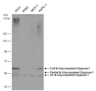
Various whole cell extracts (30 µg) were separated by 7.5% SDS-PAGE, and the membrane was blotted with Glypican 1 antibody [N3C3] diluted at 1:1500. The HRP-conjugated anti-rabbit IgG antibody was used to detect the primary antibody.Application
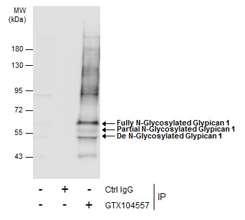
Immunoprecipitation of Glypican 1 protein from MCF-7 whole cell extracts using 5 μg of Glypican 1 antibody [N3C3] . Western blot analysis was performed using Glypican 1 antibody [N3C3] . EasyBlot anti-Rabbit IgG was used as a secondary reagent.Application
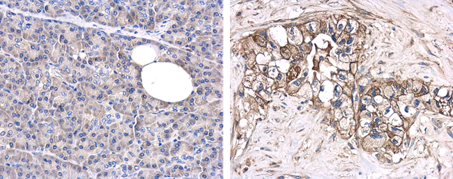
Immunohistochemical microscopy analysis of paraffin-embedded human normal pancreas (left) or pancreatic adenocarcinoma (grade II) (right) tissues using Glypican-1 antibody [N3C3] at a 1:250 dilution.Application
Untreated (–) and treated (+) A431 whole cell extracts (30 µg) were separated by 7.5% SDS-PAGE, and the membrane was blotted with Glypican 1 antibody [N3C3] diluted at 1:500. The HRP-conjugated anti-rabbit IgG antibody was used to detect the primary antibody.Application
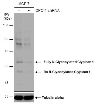
Non-transfected (–) and transfected (+) MCF-7 whole cell extracts (50 µg) were separated by 7.5% SDS-PAGE, and the membrane was blotted with Glypican 1 antibody [N3C3] diluted at 1:1500. The HRP-conjugated anti-rabbit IgG antibody was used to detect the primary antibody.Application
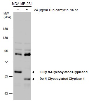
Untreated (–) and treated (+) MDA-MB-231 whole cell extracts (30 µg) were separated by 7.5% SDS-PAGE, and the membrane was blotted with Glypican 1 antibody [N3C3] diluted at 1:1500. The HRP-conjugated anti-rabbit IgG antibody was used to detect the primary antibody.Application
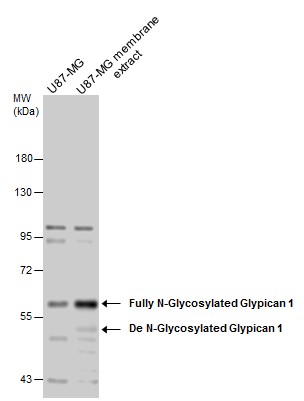
U87-MG whole cell and membrane extracts (30 µg) were separated by 7.5% SDS-PAGE, and the membrane was blotted with Glypican 1 antibody [N3C3] diluted at 1:1500. The HRP-conjugated anti-rabbit IgG antibody was used to detect the primary antibody.Application
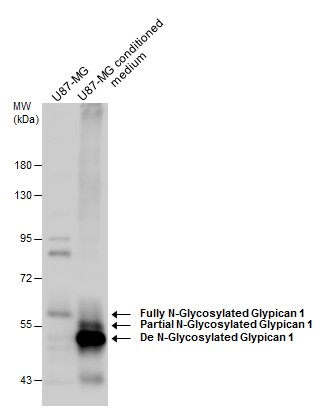
U87-MG whole cell extract and conditioned medium (30 µg) were separated by 7.5% SDS-PAGE, and the membrane was blotted with Glypican 1 antibody [N3C3] diluted at 1:1500. The HRP-conjugated anti-rabbit IgG antibody was used to detect the primary antibody.Application
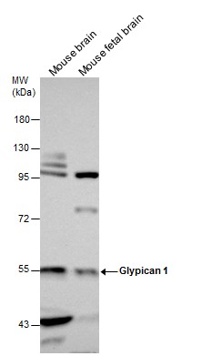
Mouse tissue extracts (50 µg) were separated by 7.5% SDS-PAGE, and the membrane was blotted with Glypican 1 antibody [N3C3] diluted at 1:1500. The HRP-conjugated anti-rabbit IgG antibody was used to detect the primary antibody.Application
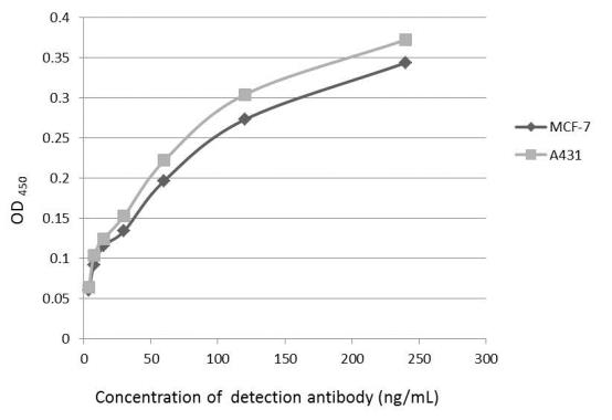
An ELISA plate is coated with MCF-7 and A431 cells. The coated cells are detected with Glypican 1 antibody at concentration ranged from 7.5 to 240 ng/mL.| Product Name | Glypican 1 antibody |
|---|---|
| Antibody Type | Primary Antibodies |
| Product description | Rabbit Polyclonal antibody to Glypican 1 (glypican 1)Cell surface heparan sulfate proteoglycans are composed of a membrane-associated protein core substituted with a variable number of heparan sulfate chains. Members of the glypican-related integral membrane proteoglycan family (GRIPS) contain a core protein anchored to the cytoplasmic membrane via a glycosyl phosphatidylinositol linkage. These proteins may play a role in the control of cell division and growth regulation. [provided by RefSeq] |
| Immunogen | Recombinant protein encompassing a sequence within the center region of human Glypican 1. The exact sequence is proprietary. |
| Clonality | Polyclonal |
|---|---|
| Isotype | IgG |
| Host Species | Rabbit |
| Tested Applications | ELISAFACSICC/IFIHC-FIHC-PIPWB |
| *Optimal dilutions/concentrations should be determined by the researcher.
: |
|
| Species Reactivity | HumanMouse |
| Predicted Reactivity | Rat, Rhesus Monkey, BovineRat (88%), Rhesus Monkey (97%), Bovine (89%) |
| Concentration | 1mg/ml |
| Purification | Affinity purified |
| Gene Synonyms | FLJ38078 antibody glypican antibody GPC1 antibody glypican proteoglycan 1 antibody glypican-1 antibody glypican 1 antibody |
|---|---|
| Molecular Weight(MW) | 62 kDa (note) |
Application
Untreated (–) and treated (+) A431 whole cell extracts (30 µg) were separated by 7.5% SDS-PAGE, and the membrane was blotted with Glypican 1 antibody [N3C3] diluted at 1:3000. The HRP-conjugated anti-rabbit IgG antibody was used to detect the primary antibody.
Application
Glypican 1 antibody [N3C3] detects Glypican 1 protein by flow cytometry analysis. Sample: A431 cell. Black: Unlabelled sample was used as a control. Red: Glypican 1 antibody [N3C3] dilution: 1:100. Acquisition of 20,000 events were collected using a Dylight 488-conjugated secondary antibody for FACS analysis.
Application
Glypican 1 antibody [N3C3] detects Glypican 1 protein at endoplasmic reticulum by immunofluorescent analysis. Sample: MCF7 cells were fixed in ice-cold MeOH for 5 min. Green: Glypican 1 protein stained by Glypican 1 antibody [N3C3] diluted at 1:400. Blue: Hoechst 33342 staining.
Application
Various whole cell extracts (30 µg) were separated by 7.5% SDS-PAGE, and the membrane was blotted with Glypican 1 antibody [N3C3] diluted at 1:1500. The HRP-conjugated anti-rabbit IgG antibody was used to detect the primary antibody.
Application
Immunoprecipitation of Glypican 1 protein from MCF-7 whole cell extracts using 5 μg of Glypican 1 antibody [N3C3] . Western blot analysis was performed using Glypican 1 antibody [N3C3] . EasyBlot anti-Rabbit IgG was used as a secondary reagent.
Application
Immunohistochemical microscopy analysis of paraffin-embedded human normal pancreas (left) or pancreatic adenocarcinoma (grade II) (right) tissues using Glypican-1 antibody [N3C3] at a 1:250 dilution.
Application
Untreated (–) and treated (+) A431 whole cell extracts (30 µg) were separated by 7.5% SDS-PAGE, and the membrane was blotted with Glypican 1 antibody [N3C3] diluted at 1:500. The HRP-conjugated anti-rabbit IgG antibody was used to detect the primary antibody.
Application
Non-transfected (–) and transfected (+) MCF-7 whole cell extracts (50 µg) were separated by 7.5% SDS-PAGE, and the membrane was blotted with Glypican 1 antibody [N3C3] diluted at 1:1500. The HRP-conjugated anti-rabbit IgG antibody was used to detect the primary antibody.
Application
Untreated (–) and treated (+) MDA-MB-231 whole cell extracts (30 µg) were separated by 7.5% SDS-PAGE, and the membrane was blotted with Glypican 1 antibody [N3C3] diluted at 1:1500. The HRP-conjugated anti-rabbit IgG antibody was used to detect the primary antibody.
Application
U87-MG whole cell and membrane extracts (30 µg) were separated by 7.5% SDS-PAGE, and the membrane was blotted with Glypican 1 antibody [N3C3] diluted at 1:1500. The HRP-conjugated anti-rabbit IgG antibody was used to detect the primary antibody.
Application
U87-MG whole cell extract and conditioned medium (30 µg) were separated by 7.5% SDS-PAGE, and the membrane was blotted with Glypican 1 antibody [N3C3] diluted at 1:1500. The HRP-conjugated anti-rabbit IgG antibody was used to detect the primary antibody.
Application
Mouse tissue extracts (50 µg) were separated by 7.5% SDS-PAGE, and the membrane was blotted with Glypican 1 antibody [N3C3] diluted at 1:1500. The HRP-conjugated anti-rabbit IgG antibody was used to detect the primary antibody.
Application
An ELISA plate is coated with MCF-7 and A431 cells. The coated cells are detected with Glypican 1 antibody at concentration ranged from 7.5 to 240 ng/mL.| Positive Control | A431 , K562 , MCF-7 , AsPC-1 , MCF-7 , A431 (500 units PNGase F for 4 hr) , MDA-MB-231 , MDA-MB-231 (24 µg/ml Tunicamycin for 16hr) , U87-MG , ME , CM , Mouse brain , mouse fetal brain |
|---|---|
| Application Notes | *Optimal dilutions/concentrations should be determined by the researcher.
: |
| Form | Liquid |
|---|---|
| Storage Instructions | Keep as concentrated solution. Aliquot and store at -20ºC or below. Avoid multiple freeze-thaw cycles. |
| Storage Buffer | 1XPBS, 20% Glycerol (pH7). 0.025% ProClin 300 was added as a preservative. |


