Application
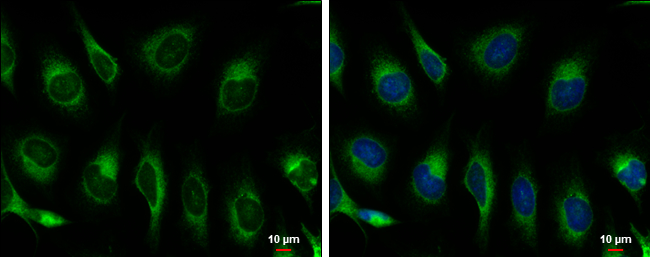
Hspa5 antibody detects Hspa5 protein at endoplasmic reticulum by immunofluorescent analysis. Sample: HeLa cells were fixed in ice-cold MeOH for 5 min. Green: Hspa5 protein stained by Hspa5 antibody diluted at 1:500. Blue: Hoechst 33342 staining. Scale bar = 10 μm.Application
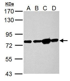
Hspa5 antibody detects Hspa5 protein by Western blot analysis. A. 50 µg Rat heart lysate/extract B. 50 µg Rat Lung lysate/extract C. 50 µg Rat Liver lysate/extract D. 50 µg Rat kidney lysate/extract 7.5 % SDS-PAGE Hspa5 antibody dilution: 1:10000Application
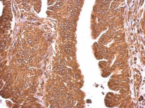
Hspa5 antibody detects Hspa5 protein at cytosol on human gastric cancer by immunohistochemical analysis. Sample: Paraffin-embedded gastric cancer. Hspa5 antibody dilution: 1:500.Application
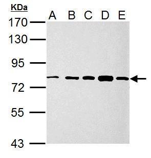
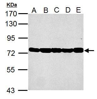
Sample (30 ug of whole cell lysate) A: 293T B: A431 C: HeLa D: HepG2 E: A375 7.5% SDS PAGE diluted at 1:10000Application
Sample (30 ug of whole cell lysate) A: NIH-3T-3 B: JC C: BCL-1 D: C2C12 E: Raw264.7 7.5% SDS PAGE diluted at 1:10000Application
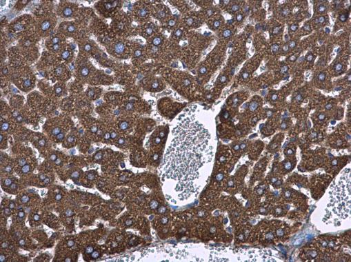
Hspa5 antibody detects Hspa5 protein at cytoplasm in mouse liver by immunohistochemical analysis. Sample: Paraffin-embedded mouse liver. Hspa5 antibody diluted at 1:500.Application
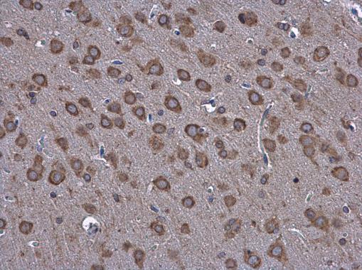
Hspa5 antibody detects Hspa5 protein at cytoplasm in rat brain by immunohistochemical analysis. Sample: Paraffin-embedded rat brain. Hspa5 antibody diluted at 1:500.| Product Name | GRP78 antibody |
|---|---|
| Antibody Type | Primary Antibodies |
| Product description | Rabbit Polyclonal antibody to GRP78Probably plays a role in facilitating the assembly of multimeric protein complexes inside the endoplasmic reticulum. Involved in the correct folding of proteins and degradation of misfolded proteins via its interaction with DNAJC10, probably to facilitate the release of DNAJC10 from its substrate. |
| Immunogen | Carrier-protein conjugated synthetic peptide encompassing a sequence within the C-terminus region of mouse Hspa5. The exact sequence is proprietary. |
| Clonality | Polyclonal |
|---|---|
| Isotype | IgG |
| Host Species | Rabbit |
| Tested Applications | ICC/IFIHC-PWB |
| *Optimal dilutions/concentrations should be determined by the researcher.
: |
|
| Species Reactivity | HumanMouseRat |
| Concentration | 1mg/ml (Please refer to the vial label for the specific concentration) |
| Purification | Affinity purified |
| Gene Synonyms | GRP-78 antibody heat shock 70kD protein 5 (glucose-regulated protein 78kD) antibody glucose regulated protein 78 kDa antibody immunoglobulin heavy chain-binding protein antibody 78 kDa glucose-regulated protein antibody heat shock 70 kDa protein 5 a |
|---|---|
| Molecular Weight(MW) | 72 kDa (note) (2) |
| Tissue Specificity | Knockdown/Knockout validation was supported by customer review data. |
| Cellular Localization | Endoplasmic reticulum lumen , Melanosome |
Application
Hspa5 antibody detects Hspa5 protein at endoplasmic reticulum by immunofluorescent analysis. Sample: HeLa cells were fixed in ice-cold MeOH for 5 min. Green: Hspa5 protein stained by Hspa5 antibody diluted at 1:500. Blue: Hoechst 33342 staining. Scale bar = 10 μm.
Application
Hspa5 antibody detects Hspa5 protein by Western blot analysis. A. 50 µg Rat heart lysate/extract B. 50 µg Rat Lung lysate/extract C. 50 µg Rat Liver lysate/extract D. 50 µg Rat kidney lysate/extract 7.5 % SDS-PAGE Hspa5 antibody dilution: 1:10000
Application
Hspa5 antibody detects Hspa5 protein at cytosol on human gastric cancer by immunohistochemical analysis. Sample: Paraffin-embedded gastric cancer. Hspa5 antibody dilution: 1:500.
Application
Sample (30 ug of whole cell lysate) A: 293T B: A431 C: HeLa D: HepG2 E: A375 7.5% SDS PAGE diluted at 1:10000
Application
Sample (30 ug of whole cell lysate) A: NIH-3T-3 B: JC C: BCL-1 D: C2C12 E: Raw264.7 7.5% SDS PAGE diluted at 1:10000
Application
Hspa5 antibody detects Hspa5 protein at cytoplasm in mouse liver by immunohistochemical analysis. Sample: Paraffin-embedded mouse liver. Hspa5 antibody diluted at 1:500.
Application
Hspa5 antibody detects Hspa5 protein at cytoplasm in rat brain by immunohistochemical analysis. Sample: Paraffin-embedded rat brain. Hspa5 antibody diluted at 1:500.| Positive Control | 293T , A431 , HeLa , HepG2 , A375 , NIH-3T-3 , JC , BCL-1 , C2C12 , Raw264.7 , rat heart , rat lung , rat liver , rat kidney |
|---|---|
| Application Notes | *Optimal dilutions/concentrations should be determined by the researcher.
: |
| Form | Liquid |
|---|---|
| Storage Instructions | Keep as concentrated solution. Aliquot and store at -20ºC or below. Avoid multiple freeze-thaw cycles. |
| Storage Buffer | 1XPBS, 0.05% Sodium Azide was added as a preservative. |


