Application
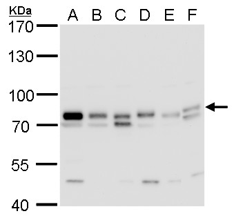
GTPBP4 antibody detects GTPBP4 protein by western blot analysis. A. 30 μg Neuro2A whole cell lysate/extract B. 30 μg GL261 whole cell lysate/extract C. 30 μg C8D30 whole cell lysate/extract D. 30 μg NIH-3T3 whole cell lysate/extract E. 30 μg BCL-1 whole cell lysate/extract F. 30 μg C2C12 whole cell lysate/extract 7.5 % SDS-PAGE GTPBP4 antibody dilution: 1:500Application
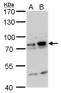
GTPBP4 antibody detects GTPBP4 protein by western blot analysis. A. 30 μg PC-12 whole cell lysate/extract B. 30 μg Rat2 whole cell lysate/extract 7.5 % SDS-PAGE GTPBP4 antibody dilution: 1:500Application
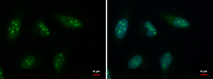
GTPBP4 antibody detects GTPBP4 protein at nucleolus by immunofluorescent analysis. Sample: HeLa cells were fixed in 4% paraformaldehyde at RT for 15 min. Green: GTPBP4 protein stained by GTPBP4 antibody diluted at 1:500. Blue: Hoechst 33342 staining.Application
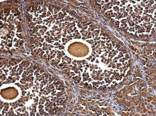
GTPBP4 antibody detects GTPBP4 protein at nucleus and nucleolus on mouse ovary by immunohistochemical analysis. Sample: Paraffin-embedded mouse ovary. GTPBP4 antibody dilution: 1:500.Application
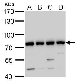
GTPBP4 antibody detects GTPBP4 protein by western blot analysis. A 293T whole cell lysate/extract B A431 C HeLa whole cell lysate/extract D HepG2 whole cell lysate/extract 7.5 % SDS-PAGE GTPBP4 antibody dilution: 1:1000| Product Name | GTPBP4 antibody |
|---|---|
| Antibody Type | Primary Antibodies |
| Product description | Rabbit Polyclonal antibody to GTPBP4 (GTP binding protein 4)GTP-binding proteins are GTPases and function as molecular switches that can flip between two states: active, when GTP is bound, and inactive, when GDP is bound. 'Active' in this context usually means that the molecule acts as a signal to trigger other events in the cell. When an extracellular ligand binds to a G-protein-linked receptor, the receptor changes its conformation and switches on the trimeric G proteins that associate with it by causing them to eject their GDP and replace it with GTP. The switch is turned off when the G protein hydrolyzes its own bound GTP, converting it back to GDP. But before that occurs, the active protein has an opportunity to diffuse away from the receptor and deliver its message for a prolonged period to its downstream target. [provided by RefSeq] |
| Immunogen | Recombinant protein encompassing a sequence within the center region of human GTPBP4. The exact sequence is proprietary. |
| Clonality | Polyclonal |
|---|---|
| Isotype | IgG |
| Host Species | Rabbit |
| Tested Applications | ICC/IFIHC-PWB |
| *Optimal dilutions/concentrations should be determined by the researcher.
: |
|
| Species Reactivity | HumanMouseRat |
| Predicted Reactivity | Chicken, BovineChicken (83%), Bovine (89%) |
| Concentration | 1mg/ml (Please refer to the vial label for the specific concentration) |
| Purification | Affinity purified |
| Gene Synonyms | NGB NOG1 GTP binding protein 4 CRFG |
|---|---|
| Target | GTPBP4 |
| Cellular Localization | Nucleus , nucleolus |
Application
GTPBP4 antibody detects GTPBP4 protein by western blot analysis. A. 30 μg Neuro2A whole cell lysate/extract B. 30 μg GL261 whole cell lysate/extract C. 30 μg C8D30 whole cell lysate/extract D. 30 μg NIH-3T3 whole cell lysate/extract E. 30 μg BCL-1 whole cell lysate/extract F. 30 μg C2C12 whole cell lysate/extract 7.5 % SDS-PAGE GTPBP4 antibody dilution: 1:500
Application
GTPBP4 antibody detects GTPBP4 protein by western blot analysis. A. 30 μg PC-12 whole cell lysate/extract B. 30 μg Rat2 whole cell lysate/extract 7.5 % SDS-PAGE GTPBP4 antibody dilution: 1:500
Application
GTPBP4 antibody detects GTPBP4 protein at nucleolus by immunofluorescent analysis. Sample: HeLa cells were fixed in 4% paraformaldehyde at RT for 15 min. Green: GTPBP4 protein stained by GTPBP4 antibody diluted at 1:500. Blue: Hoechst 33342 staining.
Application
GTPBP4 antibody detects GTPBP4 protein at nucleus and nucleolus on mouse ovary by immunohistochemical analysis. Sample: Paraffin-embedded mouse ovary. GTPBP4 antibody dilution: 1:500.
Application
GTPBP4 antibody detects GTPBP4 protein by western blot analysis. A 293T whole cell lysate/extract B A431 C HeLa whole cell lysate/extract D HepG2 whole cell lysate/extract 7.5 % SDS-PAGE GTPBP4 antibody dilution: 1:1000| Positive Control | 293T , A431 , HeLa , HepG2 , Neuro 2A , GL261 , C8D30 , NIH-3T3 , BCL-1, C2C12 , PC-12 , Rat2 |
|---|---|
| Application Notes | *Optimal dilutions/concentrations should be determined by the researcher.
: |
| Form | Liquid |
|---|---|
| Storage Instructions | Keep as concentrated solution. Aliquot and store at -20ºC or below. Avoid multiple freeze-thaw cycles. |
| Storage Buffer | 1XPBS, 20% Glycerol (pH7). 0.01% Thimerosal was added as a preservative. |


