Application
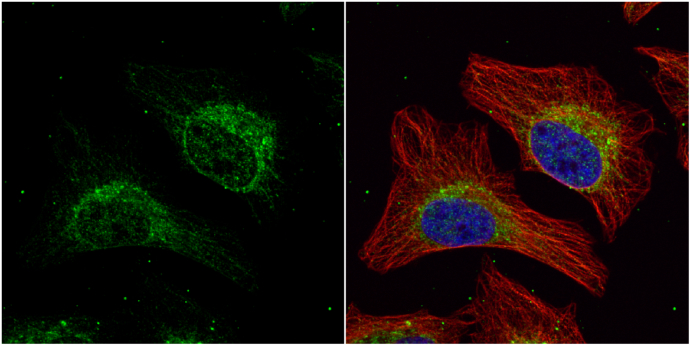
HADHA antibody [N2C1], Internal detects HADHA protein at cytoplasm by immunofluorescent analysis. Sample: HeLa cells were fixed in 4% paraformaldehyde at RT for 15 min. Green: HADHA protein stained by HADHA antibody [N2C1], Internal diluted at 1:500. Red: alpha Tubulin, a cytoskeleton marker, stained by alpha Tubulin antibody [GT114] diluted at 1:1000. Blue: Hoechst 33342 staining.Application
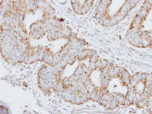
Immunohistochemical analysis of paraffin-embedded human colon carcinoma, using HADHA antibody at 1:250 dilution.Application
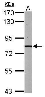
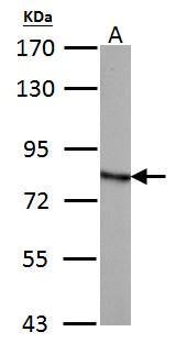
Sample (50 ug of whole cell lysate) A: mouse liver 7.5% SDS PAGE diluted at 1:1000Application
HADHA antibody [N2C1], Internal detects HADHA protein by Western blot analysis. A.30 µg PC-12 whole cell lysate/extract 7.5 % SDS-PAGE HADHA antibody [N2C1], Internal dilution: 1:500Application
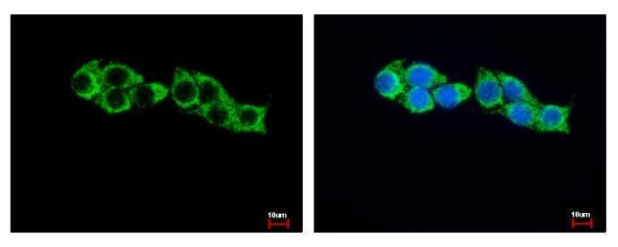
HADHA antibody [N2C1], Internal detects HADHA protein at Mitochondia by immunofluorescent analysis. Sample: HepG2 cells were fixed in -20℃ 100% MeOH for 5 min. Green: HADHA protein stained by HADHA antibody [N2C1], Internal diluted at 1:500. Blue: Hoechst 33343 staining.Application
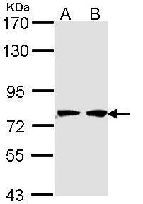
Sample (30 ug of whole cell lysate) A: 293T B: A431 7.5% SDS PAGE diluted at 1:1000| Product Name | HADHA antibody , Internal |
|---|---|
| Antibody Type | Primary Antibodies |
| Product description | Rabbit Polyclonal antibody to HADHA (hydroxyacyl-CoA dehydrogenase/3-ketoacyl-CoA thiolase/enoyl-CoA hydratase (trifunctional protein), alpha subunit)This gene encodes the alpha subunit of the mitochondrial trifunctional protein, which catalyzes the last three steps of mitochondrial beta-oxidation of long chain fatty acids. The mitochondrial membrane-bound heterocomplex is composed of four alpha and four beta subunits, with the alpha subunit catalyzing the 3-hydroxyacyl-CoA dehydrogenase and enoyl-CoA hydratase activities. Mutations in this gene result in trifunctional protein deficiency or LCHAD deficiency. The genes of the alpha and beta subunits of the mitochondrial trifunctional protein are located adjacent to each other in the human genome in a head-to-head orientation. [provided by RefSeq] |
| Immunogen | Recombinant protein encompassing a sequence within the center region of human HADHA. The exact sequence is proprietary. |
| Clonality | Polyclonal |
|---|---|
| Isotype | IgG |
| Host Species | Rabbit |
| Tested Applications | ICC/IFIHC-PWB |
| *Optimal dilutions/concentrations should be determined by the researcher.
: |
|
| Species Reactivity | HumanMouseRat |
| Predicted Reactivity | Pig, Sheep, Rhesus Monkey, BovinePig (92%), Sheep (89%), Rhesus Monkey (97%), Bovine (89%) |
| Concentration | 1mg/ml |
| Purification | Affinity purified |
| Gene Synonyms | ECHA antibody GBP antibody HADH antibody LCEH antibody LCHAD antibody MGC1728 antibody MTPA antibody TP-ALPHA antibody HADHA antibody trifunctional enzyme subunit alpha mitochondrial antibody gastrin-binding protein antibody hydroxyacyl-Coenzym |
|---|---|
| Molecular Weight(MW) | 83 kDa (note) (2) |
| Cellular Localization | Mitochondrion |
Application
HADHA antibody [N2C1], Internal detects HADHA protein at cytoplasm by immunofluorescent analysis. Sample: HeLa cells were fixed in 4% paraformaldehyde at RT for 15 min. Green: HADHA protein stained by HADHA antibody [N2C1], Internal diluted at 1:500. Red: alpha Tubulin, a cytoskeleton marker, stained by alpha Tubulin antibody [GT114] diluted at 1:1000. Blue: Hoechst 33342 staining.
Application
Immunohistochemical analysis of paraffin-embedded human colon carcinoma, using HADHA antibody at 1:250 dilution.
Application
Sample (50 ug of whole cell lysate) A: mouse liver 7.5% SDS PAGE diluted at 1:1000
Application
HADHA antibody [N2C1], Internal detects HADHA protein by Western blot analysis. A.30 µg PC-12 whole cell lysate/extract 7.5 % SDS-PAGE HADHA antibody [N2C1], Internal dilution: 1:500
Application
HADHA antibody [N2C1], Internal detects HADHA protein at Mitochondia by immunofluorescent analysis. Sample: HepG2 cells were fixed in -20℃ 100% MeOH for 5 min. Green: HADHA protein stained by HADHA antibody [N2C1], Internal diluted at 1:500. Blue: Hoechst 33343 staining.
Application
Sample (30 ug of whole cell lysate) A: 293T B: A431 7.5% SDS PAGE diluted at 1:1000| Positive Control | 293T , A431 , mouse liver , PC-12 |
|---|---|
| Application Notes | *Optimal dilutions/concentrations should be determined by the researcher.
: |
| Form | Liquid |
|---|---|
| Storage Instructions | Keep as concentrated solution. Aliquot and store at -20ºC or below. Avoid multiple freeze-thaw cycles. |
| Storage Buffer | 0.1M Tris, 0.1M Glycine, 20% Glycerol (pH7). 0.01% Thimerosal was added as a preservative. |


