Application
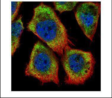
Confocal immunofluorescence analysis (Olympus FV10i) of methanol-fixed A431, using HAX1 antibody (Green) at 1:500 dilution. Alpha-tubulin filaments were labeled with (Red) at 1:2000.Application
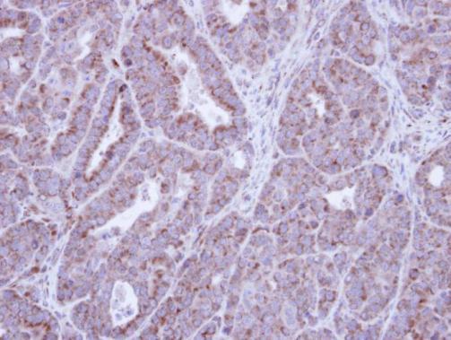
Immunohistochemical analysis of paraffin-embedded NCIN87 Xenograft , using HAX1 antibody at 1:100 dilution.Application
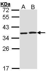
Sample (30 µg of whole cell lysate) A: Molt-4 B: Raji 12% SDS PAGE diluted at 1:1000 The HRP-conjugated anti-rabbit IgG antibody was used to detect the primary antibody.Application
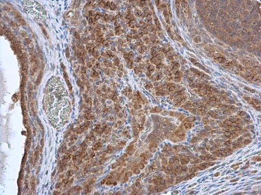
HAX1 antibody detects HAX1 protein at cytoplasm in rat ovary by immunohistochemical analysis. Sample: Paraffin-embedded rat ovary. HAX1 antibody diluted at 1:500.Application
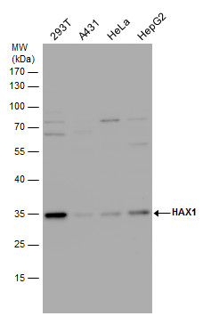
HAX1 antibody detects HAX1 protein by western blot analysis. Various whole cell extracts (30 µg) were separated by 12% SDS-PAGE, and the membrane was blotted with HAX1 antibody diluted by 1:1000. The HRP-conjugated anti-rabbit IgG antibody was used to detect the primary antibody.| Product Name | HAX1 antibody |
|---|---|
| Antibody Type | Primary Antibodies |
| Product description | Rabbit Polyclonal antibody to HAX1 (HCLS1 associated protein X-1)The protein encoded by this gene is known to associate with hematopoietic cell-specific Lyn substrate 1, a substrate of Src family tyrosine kinases. It also interacts with the product of the polycystic kidney disease 2 gene, mutations in which are associated with autosomal-dominant polycystic kidney disease, and with the F-actin-binding protein, cortactin. It was earlier thought that this gene product is mainly localized in the mitochondria, however, recent studies indicate it to be localized in the cell body. Mutations in this gene result in autosomal recessive severe congenital neutropenia, also known as Kostmann disease. Two transcript variants encoding different isoforms have been found for this gene. [provided by RefSeq] |
| Immunogen | Recombinant protein encompassing a sequence within the center region of human HAX1. The exact sequence is proprietary. |
| Clonality | Polyclonal |
|---|---|
| Isotype | IgG |
| Host Species | Rabbit |
| Tested Applications | ICC/IFIHC-PWB |
| *Optimal dilutions/concentrations should be determined by the researcher.
: |
|
| Species Reactivity | HumanRat |
| Predicted Reactivity | Rhesus Monkey, BovineRhesus Monkey (97%), Bovine (84%) |
| Concentration | 1mg/ml (Please refer to the vial label for the specific concentration) |
| Purification | Affinity purified |
| Gene Synonyms | FLJ17042 antibody FLJ18492 antibody FLJ93803 antibody HCLSBP1 antibody HS1BP1 antibody SCN3 antibody HAX1 antibody HAX-1 antibody HCLS1-associated protein X-1 antibody HSP1BP-1 antibody HS1-binding protein 1 antibody HS1 binding protein antibody |
|---|---|
| Molecular Weight(MW) | 32 kDa (note) |
| Cellular Localization | Mitochondrion , Endoplasmic reticulum , Nucleus membrane |
Application
Confocal immunofluorescence analysis (Olympus FV10i) of methanol-fixed A431, using HAX1 antibody (Green) at 1:500 dilution. Alpha-tubulin filaments were labeled with (Red) at 1:2000.
Application
Immunohistochemical analysis of paraffin-embedded NCIN87 Xenograft , using HAX1 antibody at 1:100 dilution.
Application
Sample (30 µg of whole cell lysate) A: Molt-4 B: Raji 12% SDS PAGE diluted at 1:1000 The HRP-conjugated anti-rabbit IgG antibody was used to detect the primary antibody.
Application
HAX1 antibody detects HAX1 protein at cytoplasm in rat ovary by immunohistochemical analysis. Sample: Paraffin-embedded rat ovary. HAX1 antibody diluted at 1:500.
Application
HAX1 antibody detects HAX1 protein by western blot analysis. Various whole cell extracts (30 µg) were separated by 12% SDS-PAGE, and the membrane was blotted with HAX1 antibody diluted by 1:1000. The HRP-conjugated anti-rabbit IgG antibody was used to detect the primary antibody.| Positive Control | Molt-4 , Raji , 293T , A431 , HeLa , HepG2 |
|---|---|
| Application Notes | *Optimal dilutions/concentrations should be determined by the researcher.
: |
| Form | Liquid |
|---|---|
| Storage Instructions | Keep as concentrated solution. Aliquot and store at -20ºC or below. Avoid multiple freeze-thaw cycles. |
| Storage Buffer | 1XPBS, 10% Glycerol (pH7). 0.01% Thimerosal was added as a preservative. |


