Application
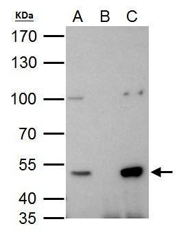
HDAC3 antibody immunoprecipitates HDAC3 protein in IP experiments. IP Sample: 293T whole cell lysate/extract A. 40 μg 293T whole cell lysate/extract B. Control with 2 μg of preimmune rabbit IgG C. Immunoprecipitation of HDAC3 protein by 2 μg of HDAC3 antibody 7.5% SDS-PAGE The immunoprecipitated HDAC3 protein was detected by HDAC3 antibody diluted at 1:1000. EasyBlot anti-rabbit IgG was used as a secondary reagent.Application
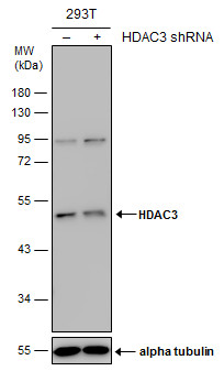
Non-transfected (–) and transfected (+) 293T whole cell extracts (30 µg) were separated by 10% SDS-PAGE, and the membrane was blotted with HDAC3 antibody diluted at 1:500. The HRP-conjugated anti-rabbit IgG antibody was used to detect the primary antibody.Application
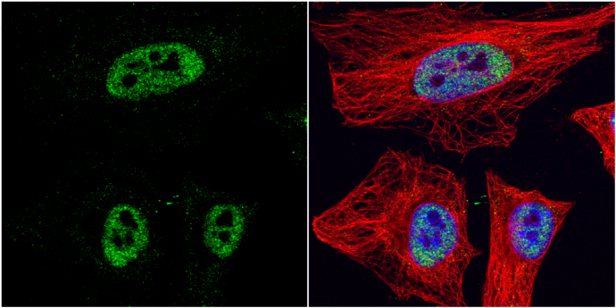
HDAC3 antibody detects HDAC3 protein at nucleus by immunofluorescent analysis. Sample: HeLa cells were fixed in 4% paraformaldehyde at RT for 15 min. Green: HDAC3 protein stained by HDAC3 antibody diluted at 1:1000. Red: alpha Tubulin, a cytoskeleton marker, stained by alpha Tubulin antibody [GT114] diluted at 1:1000. Blue: Hoechst 33342 staining.Application
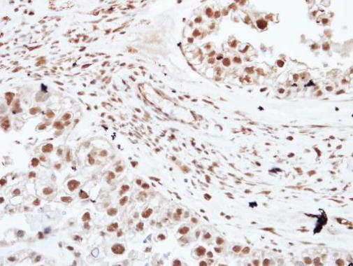
Immunohistochemical analysis of paraffin-embedded human ovarian cancer, using HDAC3 antibody at 1:250 dilution.Application
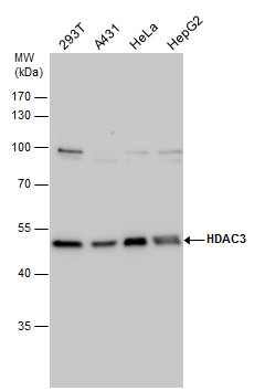
HDAC3 antibody detects HDAC3 protein by western blot analysis. Various whole cell extracts (30 µg) were separated by 10% SDS-PAGE, and the membrane was blotted with HDAC3 antibody diluted by 1:1000. The HRP-conjugated anti-rabbit IgG antibody was used to detect the primary antibody.Application
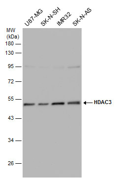
Various whole cell extracts (30 µg) were separated by 10% SDS-PAGE, and the membrane was blotted with HDAC3 antibody diluted at 1:500. The HRP-conjugated anti-rabbit IgG antibody was used to detect the primary antibody.Application
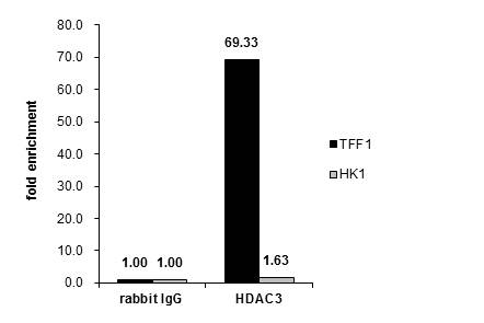
Cross-linked ChIP was performed with MCF-7 chromatin extract and 5 μg of either control rabbit IgG or anti-HDAC3 antibody. The precipitated DNA was detected by PCR with primer set targeting to TFF1 or HK1.WB
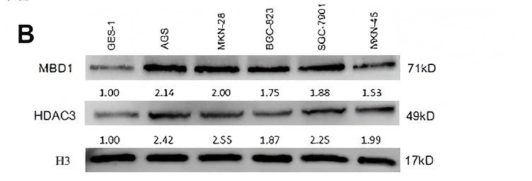
(B) MBD/HDAC3 levels of several GC cell lines were measured by western blot.IHC
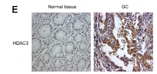
(E) The representative images of HDAC3 in GC tissue versus normal tissue by immunohistochemistry.IF
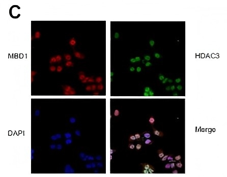
(C) Immunofluorescence staining showed that MBD1 and HDAC3 co-localized in nuclei of SGC-7901 cells.WB
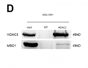
(D) IP assay showed that MBD1 directly bound to HDAC3 at the protein level.| Product Name | HDAC3 antibody |
|---|---|
| Antibody Type | Primary Antibodies |
| Product description | Rabbit Polyclonal antibody to HDAC3 (histone deacetylase 3)Histones play a critical role in transcriptional regulation, cell cycle progression, and developmental events. Histone acetylation/deacetylation alters chromosome structure and affects transcription factor access to DNA. The protein encoded by this gene belongs to the histone deacetylase/acuc/apha family. It has histone deacetylase activity and represses transcription when tethered to a promoter. It may participate in the regulation of transcription through its binding with the zinc-finger transcription factor YY1. This protein can also down-regulate p53 function and thus modulate cell growth and apoptosis. This gene is regarded as a potential tumor suppressor gene. [provided by RefSeq] |
| Immunogen | Recombinant protein encompassing a sequence within the center region of human HDAC3. The exact sequence is proprietary. |
| Isotype | IgG |
|---|---|
| Host Species | Rabbit |
| Tested Applications | ChIPICC/IFIFIHCIHC-PIPWB |
| *Optimal dilutions/concentrations should be determined by the researcher. | |
| Species Reactivity | HumanMouseRat |
| Predicted Reactivity | Zebrafish, Pig, Chicken, Rhesus Monkey, BovineZebrafish (89%), Pig (100%), Chicken (95%), Rhesus Monkey (100%), Bovine (100%) |
| Concentration | 1mg/ |
| Purification | Affinity purified |
| Gene Synonyms | HD3 antibody, RPD3 antibody, RPD3-2 antibody, HDAC3 antibody, SMAP45 antibody, histone deacetylase 3 antibody |
|---|---|
| Molecular Weight(MW) | 49 kDa (note) (2) |
| Cellular Localization | Nucleus |
Application
HDAC3 antibody immunoprecipitates HDAC3 protein in IP experiments. IP Sample: 293T whole cell lysate/extract A. 40 μg 293T whole cell lysate/extract B. Control with 2 μg of preimmune rabbit IgG C. Immunoprecipitation of HDAC3 protein by 2 μg of HDAC3 antibody 7.5% SDS-PAGE The immunoprecipitated HDAC3 protein was detected by HDAC3 antibody diluted at 1:1000. EasyBlot anti-rabbit IgG was used as a secondary reagent.
Application
Non-transfected (–) and transfected (+) 293T whole cell extracts (30 µg) were separated by 10% SDS-PAGE, and the membrane was blotted with HDAC3 antibody diluted at 1:500. The HRP-conjugated anti-rabbit IgG antibody was used to detect the primary antibody.
Application
HDAC3 antibody detects HDAC3 protein at nucleus by immunofluorescent analysis. Sample: HeLa cells were fixed in 4% paraformaldehyde at RT for 15 min. Green: HDAC3 protein stained by HDAC3 antibody diluted at 1:1000. Red: alpha Tubulin, a cytoskeleton marker, stained by alpha Tubulin antibody [GT114] diluted at 1:1000. Blue: Hoechst 33342 staining.
Application
Immunohistochemical analysis of paraffin-embedded human ovarian cancer, using HDAC3 antibody at 1:250 dilution.
Application
HDAC3 antibody detects HDAC3 protein by western blot analysis. Various whole cell extracts (30 µg) were separated by 10% SDS-PAGE, and the membrane was blotted with HDAC3 antibody diluted by 1:1000. The HRP-conjugated anti-rabbit IgG antibody was used to detect the primary antibody.
Application
Various whole cell extracts (30 µg) were separated by 10% SDS-PAGE, and the membrane was blotted with HDAC3 antibody diluted at 1:500. The HRP-conjugated anti-rabbit IgG antibody was used to detect the primary antibody.
Application
Cross-linked ChIP was performed with MCF-7 chromatin extract and 5 μg of either control rabbit IgG or anti-HDAC3 antibody. The precipitated DNA was detected by PCR with primer set targeting to TFF1 or HK1.
WB
(B) MBD/HDAC3 levels of several GC cell lines were measured by western blot.
IHC
(E) The representative images of HDAC3 in GC tissue versus normal tissue by immunohistochemistry.
IF
(C) Immunofluorescence staining showed that MBD1 and HDAC3 co-localized in nuclei of SGC-7901 cells.
WB
(D) IP assay showed that MBD1 directly bound to HDAC3 at the protein level.| Positive Control | 293T , A431 , HeLa , HepG2 , U87-MG , SK-N-SH , IMR32 , SK-N-AS , *NB2a |
|---|---|
| Application Notes | *Optimal dilutions/concentrations should be determined by the researcher. |
| Form | Liquid |
|---|---|
| Storage Instructions | Keep as concentrated solution. Aliquot and store at -20ºC or below. Avoid multiple freeze-thaw cycles. |
| Storage Buffer | 1XPBS, 1% BSA, 20% Glycerol (pH7). 0.01% Thimerosal was added as a preservative. |


