Application
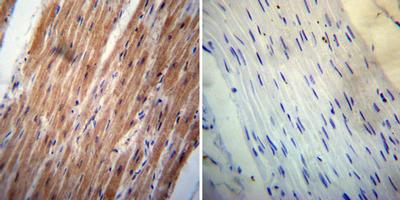
IHC-P analysis of human heart tissue with (left) or without (right) ErbB4 antibody [HFR1] at a dilution of 1:200. To expose target proteins, heat induced antigen retrieval was performed using 10mM sodium citrate (pH6.0) buffer, microwaved for 8-15 minutes.Application
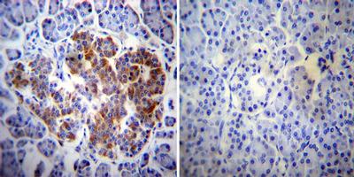
IHC-P analysis of human pancreas tissue with (left) or without (right) ErbB4 antibody [HFR1] at a dilution of 1:200. To expose target proteins, heat induced antigen retrieval was performed using 10mM sodium citrate (pH6.0) buffer, microwaved for 8-15 minutes.Application
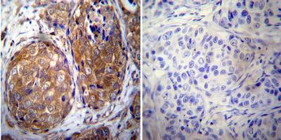
IHC-P analysis of human breast carcinoma with (left) or without (right) ErbB4 antibody [HFR1] at a dilution of 1:20. To expose target proteins, heat induced antigen retrieval was performed using 10mM sodium citrate (pH6.0) buffer, microwaved for 8-15 minutes.Application
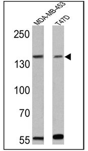
WB analysis of MDA-MB-453 and T47D lysates (25 ug per lane) using ErbB4 antibody [HFR1] at a dilution of 1:300.Application
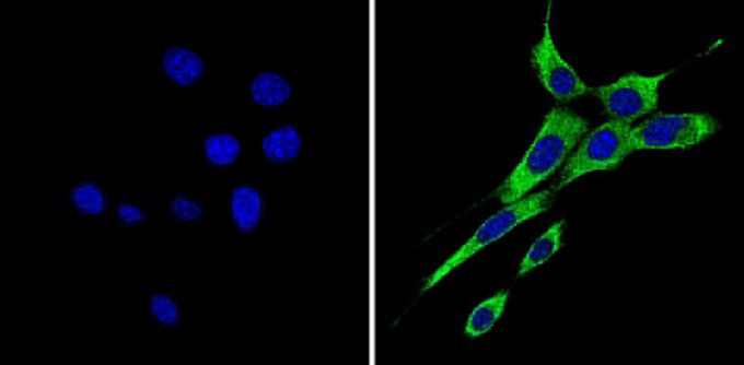
ICC/IF analysis of NIH-3T3 cells with (right) or without (left) ErbB4 antibody [HFR1] at a dilution of 1:50 (green). Nuclei were stained with DAPI (blue). Images were taken at a magnification of 60x.Application
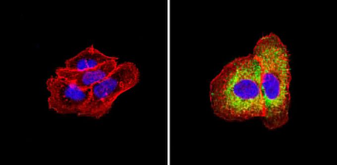
ICC/IF analysis of A431 cells with (right) or without (left) ErbB4 antibody [HFR1] at a dilution of 1:50 (green). Actin was stained using Alexa Fluor 554 (red) and nuclei were stained with DAPI (blue). Images were taken at a magnification of 60x.Application
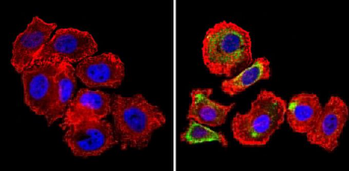
ICC/IF analysis of SK-BR-3 cells with (right) or without (left) ErbB4 antibody [HFR1] at a dilution of 1:50 (green). Actin was stained using Alexa Fluor 554 (red) and nuclei were stained with DAPI (blue). Images were taken at a magnification of 60x.Application
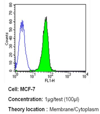
FACS analysis of MCF-7 cells with ErbB4 antibody [HFR1] at a dilution of 1ug/test (green) or an isotype control (blue).Application
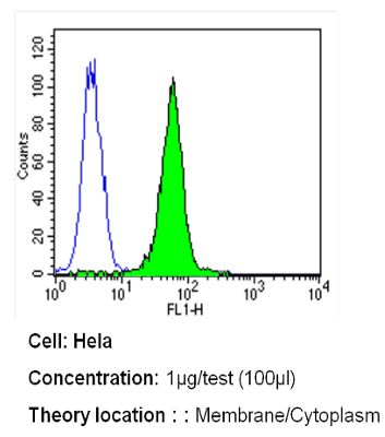
FACS analysis of HeLa cells with ErbB4 antibody [HFR1] at a dilution of 1ug/test (green) or an isotype control (blue).Application
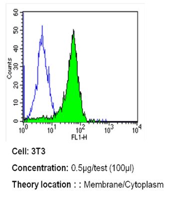
FACS analysis of NIH-3T3 cells with ErbB4 antibody [HFR1] at a dilution of 0.5ug/test (green) or an isotype control (blue).| Product Name | Her4 / ErbB4 antibody |
|---|---|
| Antibody Type | Primary Antibodies |
| Product description | Mouse monoclonal antibody [HFR1] to ErbB4This gene is a member of the Tyr protein kinase family and the epidermal growth factor receptor subfamily. It encodes a single-pass type I membrane protein with multiple cysteine rich domains, a transmembrane domain, a tyrosine kinase domain, a phosphotidylinositol-3 kinase binding site and a PDZ domain binding motif. The protein binds to and is activated by neuregulins and other factors and induces a variety of cellular responses including mitogenesis and differentiation. Multiple proteolytic events allow for the release of a cytoplasmic fragment and an extracellular fragment. Mutations in this gene have been associated with cancer. Alternatively spliced variants which encode different protein isoforms have been described; however, not all variants have been fully characterized. [provided by RefSeq, Jul 2008] |
| Immunogen | Synthetic Peptide: C R(1250) S T L Q H P D Y L Q E Y S T(1264) |
| Clonality | Monoclonal |
|---|---|
| Isotype | IgG2 |
| Clone Number | HFR1 |
| Host Species | Mouse |
| Tested Applications | FACSICC/IFIHCIHC-PIPWB |
| IHC-P: 6 - 8 µg/mL: |
|
| Species Reactivity | HumanMouse |
| Concentration | 1mg/ml (Please refer to the vial label for the specific concentration) |
| Purification | Protein A |
| Gene Synonyms | v-erb-a erythroblastic leukemia viral oncogene homolog 4 (avian) ERBB4 p180erbB4 HER4 |
|---|---|
| Target | ErbB4 |
Application
IHC-P analysis of human heart tissue with (left) or without (right) ErbB4 antibody [HFR1] at a dilution of 1:200. To expose target proteins, heat induced antigen retrieval was performed using 10mM sodium citrate (pH6.0) buffer, microwaved for 8-15 minutes.
Application
IHC-P analysis of human pancreas tissue with (left) or without (right) ErbB4 antibody [HFR1] at a dilution of 1:200. To expose target proteins, heat induced antigen retrieval was performed using 10mM sodium citrate (pH6.0) buffer, microwaved for 8-15 minutes.
Application
IHC-P analysis of human breast carcinoma with (left) or without (right) ErbB4 antibody [HFR1] at a dilution of 1:20. To expose target proteins, heat induced antigen retrieval was performed using 10mM sodium citrate (pH6.0) buffer, microwaved for 8-15 minutes.
Application
WB analysis of MDA-MB-453 and T47D lysates (25 ug per lane) using ErbB4 antibody [HFR1] at a dilution of 1:300.
Application
ICC/IF analysis of NIH-3T3 cells with (right) or without (left) ErbB4 antibody [HFR1] at a dilution of 1:50 (green). Nuclei were stained with DAPI (blue). Images were taken at a magnification of 60x.
Application
ICC/IF analysis of A431 cells with (right) or without (left) ErbB4 antibody [HFR1] at a dilution of 1:50 (green). Actin was stained using Alexa Fluor 554 (red) and nuclei were stained with DAPI (blue). Images were taken at a magnification of 60x.
Application
ICC/IF analysis of SK-BR-3 cells with (right) or without (left) ErbB4 antibody [HFR1] at a dilution of 1:50 (green). Actin was stained using Alexa Fluor 554 (red) and nuclei were stained with DAPI (blue). Images were taken at a magnification of 60x.
Application
FACS analysis of MCF-7 cells with ErbB4 antibody [HFR1] at a dilution of 1ug/test (green) or an isotype control (blue).
Application
FACS analysis of HeLa cells with ErbB4 antibody [HFR1] at a dilution of 1ug/test (green) or an isotype control (blue).
Application
FACS analysis of NIH-3T3 cells with ErbB4 antibody [HFR1] at a dilution of 0.5ug/test (green) or an isotype control (blue).| Application Notes | IHC-P: 6 - 8 µg/mL: |
|---|
| Form | Liquid |
|---|---|
| Storage Instructions | -20° C, Avoid Freeze/Thaw Cycles |
| Storage Buffer | PBS containing 0.05 % sodium azide and 1 mg/ml BSA |


