Application
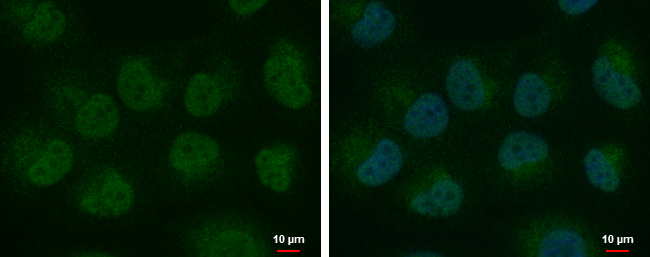
HEY1 antibody detects HEY1 protein at nucleus and cytoplasm by immunofluorescent analysis. Sample: H1299 cells were fixed in 4% paraformaldehyde at RT for 15 min. Green: HEY1 protein stained by HEY1 antibody diluted at 1:1000. Blue: Hoechst 33342 staining. Scale bar = 10 μm.Application
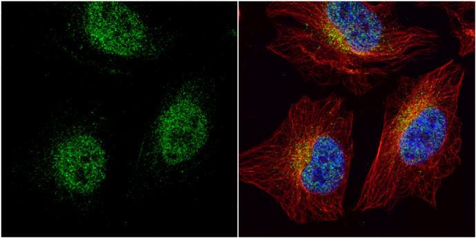
HEY1 antibody detects HEY1 protein at cytoplasm and nucleus by immunofluorescent analysis. Sample: HeLa cells were fixed in 4% paraformaldehyde at RT for 15 min. Green: HEY1 protein stained by HEY1 antibody diluted at 1:2000. Red: alpha Tubulin, a cytoskeleton marker, stained by alpha Tubulin antibody [GT114] diluted at 1:1000. Blue: Hoechst 33342 staining.Application
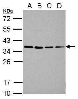
Sample (30 µg of whole cell lysate) A: A549 B: H1299 C: HCT116 D: MCF-7 12% SDS PAGE diluted at 1:1000 The HRP-conjugated anti-rabbit IgG antibody was used to detect the primary antibody.| Product Name | HEY1 antibody |
|---|---|
| Antibody Type | Primary Antibodies |
| Product description | Rabbit Polyclonal antibody to HEY1 (hairy/enhancer-of-split related with YRPW motif 1)This gene encodes a nuclear protein belonging to the hairy and enhancer of split-related (HESR) family of basic helix-loop-helix (bHLH)-type transcriptional repressors. Expression of this gene is induced by the Notch and c-Jun signal transduction pathways. Two similar and redundant genes in mouse are required for embryonic cardiovascular development, and are also implicated in neurogenesis and somitogenesis. Alternative splicing results in multiple transcript variants. [provided by RefSeq] |
| Immunogen | Recombinant protein encompassing a sequence within the center region of human HEY1. The exact sequence is proprietary. |
| Clonality | Polyclonal |
|---|---|
| Isotype | IgG |
| Host Species | Rabbit |
| Tested Applications | ICC/IFWB |
| *Optimal dilutions/concentrations should be determined by the researcher.
: |
|
| Species Reactivity | Human |
| Predicted Reactivity | Mouse, Rat, Xenopus laevis, Dog, BovineMouse (96%), Rat (89%), Xenopus laevis (81%), Dog (94%), Bovine (93%) |
| Concentration | 1mg/ml (Please refer to the vial label for the specific concentration) |
| Purification | Affinity purified |
| Gene Synonyms | BHLHb31 antibody CHF2 antibody HERP2 antibody HESR1 antibody HRT-1 antibody MGC1274 antibody OAF1 antibody HEY1 antibody HES-related repressor protein 1 antibody basic helix-loop-helix protein OAF1 antibody cardiovascular helix-loop-helix factor |
|---|---|
| Molecular Weight(MW) | 33 kDa (note) (2) |
| Cellular Localization | Nucleus |
Application
HEY1 antibody detects HEY1 protein at nucleus and cytoplasm by immunofluorescent analysis. Sample: H1299 cells were fixed in 4% paraformaldehyde at RT for 15 min. Green: HEY1 protein stained by HEY1 antibody diluted at 1:1000. Blue: Hoechst 33342 staining. Scale bar = 10 μm.
Application
HEY1 antibody detects HEY1 protein at cytoplasm and nucleus by immunofluorescent analysis. Sample: HeLa cells were fixed in 4% paraformaldehyde at RT for 15 min. Green: HEY1 protein stained by HEY1 antibody diluted at 1:2000. Red: alpha Tubulin, a cytoskeleton marker, stained by alpha Tubulin antibody [GT114] diluted at 1:1000. Blue: Hoechst 33342 staining.
Application
Sample (30 µg of whole cell lysate) A: A549 B: H1299 C: HCT116 D: MCF-7 12% SDS PAGE diluted at 1:1000 The HRP-conjugated anti-rabbit IgG antibody was used to detect the primary antibody.| Positive Control | A549 , H1299 , HCT116 , MCF-7 |
|---|---|
| Application Notes | *Optimal dilutions/concentrations should be determined by the researcher.
: |
| Form | Liquid |
|---|---|
| Storage Instructions | Keep as concentrated solution. Aliquot and store at -20ºC or below. Avoid multiple freeze-thaw cycles. |
| Storage Buffer | 1XPBS, 20% Glycerol (pH7). 0.01% Thimerosal was added as a preservative. |


