Application
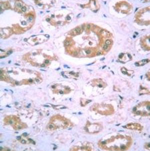
Immunohistochemical staining of human kidney tissue using HIF1 alpha mouse monoclonal antibody [H1alpha67].Application
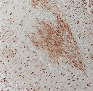
Immunohistochemical staining of human lung tissue using HIF1 alpha mouse monoclonal antibody [H1alpha67].Application
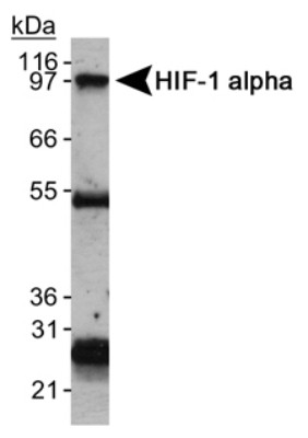
Immunoprecipitation of HIF1 alpha protein using HIF1 alpha mouse monoclonal antibody [H1alpha67]. The heavy and light chains are also detected.Application
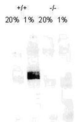
Western Blot: HIF-1 alpha Antibody (H1alpha67) - On day 1, MEF cells (+/+,-/-), were grown on 15 cm dish (2x10 to the 6th cells). On day 2, cells were exposed to hypoxia for 4hrs. Cells were washed with ice cold PBS twice and whole cell protein was extracted with RIPA buffer fortified with protease. Upon quantification, 100ug of protein was fractionated on 7% polyacralymide gel. Gel was transferred overnight onto nitrocellulose membrane. The membrane was probed with HIF-1 alpha monoclonal antibody at a 1:500 dilution . The secondary antibody was conjugated with HRP and was used at a 1:2500 dilutionApplication
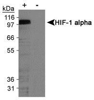
WB analysis of cobalt chloride treated and untreated COS-7 nuclear extracts using HIF1 alpha antibody [H1alpha67].Application
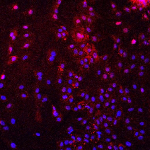
ICC/IF analysis of pig endothelial cells under hipoxia condition using HIF1 alpha antibody [H1alpha67] (red) and DAPI.Application
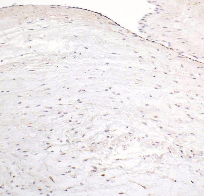
IHC-P analysis of pig tissue (brown) using HIF1 alpha antibody [H1alpha67].| Product Name | HIF1 alpha antibody |
|---|---|
| Antibody Type | Primary Antibodies |
| Product description | Mouse monoclonal antibody [H1alpha67] to HIF1 alphaHypoxia contributes significantly to the pathophysiology of major categories of human disease, including myocardial and cerebral ischemia, cancer, pulmonary hypertension, congenital heart disease and chronic obstructive pulmonary disease. |
| Immunogen | Amino acids 432-528 of human HIF1 alpha |
| Clonality | Monoclonal |
|---|---|
| Isotype | IgG2 |
| Clone Number | H1alpha67 |
| Host Species | Mouse |
| Tested Applications | ELISAFACSGSAICC/IFIHCIHC-PIPWBChIP |
| For WB: Use at a dilution of 1:500-1:1000: |
|
| Species Reactivity | HumanMouseDogPigRatFerretPrimateSheepBovineAvian |
| Concentration | 1mg/ml |
| Purification | Protein A |
| Gene Synonyms | HIF1 alpha HIF-1alpha HIF1A MOP1 PASD8 HIF1-ALPHA 603348 HIF1ALPHA Q16665 3091 HIF 1alpha |
|---|---|
| Target | HIF1 alpha |
| Tissue Specificity | This antibody is specific to HIF1 alpha |
| Cellular Localization | HIF1 is a nuclear protein that activates gene transcription in response to reduced cellular O2 concentration. Activates transcription of EPO, VEGF, iNOS, heme oxygenase 1 and other critical intracellular responses to hypoxia. |
Application
Immunohistochemical staining of human kidney tissue using HIF1 alpha mouse monoclonal antibody [H1alpha67].
Application
Immunohistochemical staining of human lung tissue using HIF1 alpha mouse monoclonal antibody [H1alpha67].
Application
Immunoprecipitation of HIF1 alpha protein using HIF1 alpha mouse monoclonal antibody [H1alpha67]. The heavy and light chains are also detected.
Application
Western Blot: HIF-1 alpha Antibody (H1alpha67) - On day 1, MEF cells (+/+,-/-), were grown on 15 cm dish (2x10 to the 6th cells). On day 2, cells were exposed to hypoxia for 4hrs. Cells were washed with ice cold PBS twice and whole cell protein was extracted with RIPA buffer fortified with protease. Upon quantification, 100ug of protein was fractionated on 7% polyacralymide gel. Gel was transferred overnight onto nitrocellulose membrane. The membrane was probed with HIF-1 alpha monoclonal antibody at a 1:500 dilution . The secondary antibody was conjugated with HRP and was used at a 1:2500 dilution
Application
WB analysis of cobalt chloride treated and untreated COS-7 nuclear extracts using HIF1 alpha antibody [H1alpha67].
Application
ICC/IF analysis of pig endothelial cells under hipoxia condition using HIF1 alpha antibody [H1alpha67] (red) and DAPI.
Application
IHC-P analysis of pig tissue (brown) using HIF1 alpha antibody [H1alpha67].| Application Notes | For WB: Use at a dilution of 1:500-1:1000: |
|---|
| Form | Liquid |
|---|---|
| Storage Instructions | Keep as concentrated solution, aliquot and store at 4°C. |
| Storage Buffer | Tris-glycine, 150mM NaCl pH 7.5 with 0.05% Sodium Azide |


